Download
1 / 47
490 likes | 723 Vues
SHOCK C omplications & Approach to Patient. Dr.Mohammed Sharique Ahmed Quadri Assistant Prof.Physiology Almaarefa College. Complications of Shock. Acute respiratory distress syndrome Acute renal failure Gastrointestinal complications Disseminated intravascular coagulation

E N D
SHOCKComplications &Approach to Patient Dr.MohammedSharique Ahmed Quadri Assistant Prof.Physiology Almaarefa College
Complications of Shock • Acute respiratory distress syndrome • Acute renal failure • Gastrointestinal complications • Disseminated intravascular coagulation • Multiple organ dysfunction syndrome
Acute Respiratory Distress Syndrome (ARDS) • Potentially life threatening form of lung injury • Characterized by • Severe dyspnea of rapid onset • Respiratory rate increases • Profound hypoxemia (cyanosis) refractory to supplemental oxygen • Results from greatly reduced diffusion of gases across the thickened alveolar membranes. • Pulmonary infiltration ( x-ray chest) • Exact cause is unknown
Pathophysiology of (ARDS) Activation of neutrophils Accumulates in pulmonary vasculature cytokines Leakage of fluid and plasma proteins in alveolar spaces Injury to endothelial cells • Atelectasis • Impaired gas exchange • Decrease compliance (stiffness) • Decrease surfactant
Clinical features OF ARDS • Tachypnea, tachycardia, hypoxia, and respiratory alkalosis are typical early clinical manifestations • Usually followed by the appearance of diffuse pulmonary infiltrates and respiratory failure within 48 hours.
Acute Renal Failure (ARF) • Renal vasoconstriction cuts off urine production • Results in Acute renal failure • Continued vasoconstriction cuts off renal oxygen supply • Renal tubular cells die leading to • Acute tubular necrosis
Acute Renal Failure (ARF) • Frequent monitoring of urine out put provided a means of assesing renal blood flow.(urine out put of 20 ml/hr or less indicate impaired renal perfusion) • Serum creatinineand blood urea nitrogen levels provided valuable information regarding renal status
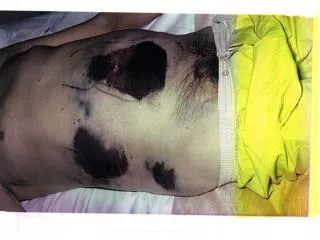
G.I Complication • Constriction of vessels supplying GIT for redistribution of blood flow • Severe Decrease mucosal perfusion • GIT ulceration • Bleeding
Disseminated IntravascularCoagulation (DIC) coagulation pathways activated clots in platelets many and small clotting blood proteins vessels used up ORAGAN FAILURE microinfarcts, bleeding ischemia problems
Multiple Organ Dysfunction Syndrome (MODS) • The most frequent cause of death in the noncoronary intensive care unit • Affects multiple organ system (kidney, heart lungs, liver & brain. • Mortality rates vary from 30% to 100% • Pathogenesis not clearly understood
Major risk factor for development of MODS are • Severe trauma • Sepsis • Prolonged periods of hypotension • Hepatic dysfunction • Infarcted bowel • Advanced age • Alcohol abuse
Assess Intervene RE-assess Seek help Avoid over reliance on invasive haemodynamic monitoring Pulse rate Capillary fill time temperature Blood pressure Level of consciousness Blood-gas estimation
Practically Speaking…. • Keep eye on these patients • Frequent vitals signs: • Monitor success of therapies • Watch for decompensated shock • Let your nurses know that these patients are sick!
Approach to the Patient in Shock • ABCs • Cardiorespiratory monitor • Pulse oximetry • Supplemental oxygen • IV access • ABG, labs • Foley catheter • Vital signs
Diagnosis • Physical exam (Vital Signs, mental status, skin color, temperature, pulses, etc.) • Surveillance for Infectious source • Labs: • CBC • Chemistries ( urea, creatinin ,etc) • Lactate • Coagulation studies • Cultures • ABG ( arterial blood gas analysis)
Further Evaluation • CVP( central venous pressure)and PCWP(pulmonary capillary wedge pressure) • CT of head/sinuses • Lumbar puncture • Wound cultures • Abdominal/pelvic CT or USG • Cortisol level • Fibrinogen, FDPs(fibrin degradation product), D-dimer
History Recent illness Fever Chest pain, SOB Abdominal pain Comorbidities Medications Toxins/Ingestions Recent hospitalization or surgery Baseline mental status Physical examination Vital Signs CNS – mental status Skin – color, temp, rashes, sores CV – JVP, heart sounds Resp – lung sounds, RR, oxygen sat, ABG GI – tenderness , rigidity, guarding, rebound Renal – urine output Approach to the Patient in Shock
Is This Patient in Shock? • Patient looks ill • Altered mental status • Skin cool and mottled or hot and flushed • Weak or absent peripheral pulses • SBP <110 • Tachycardia Yes! These are all signs and symptoms of shock
Shock • Do you remember how to quickly estimate blood pressure by pulse? 60 • by palpating a pulse, • you know SBP is at • least this number 70 80 90
Goals of Treatment • ABCDE • Airway • control work of Breathing • optimize Circulation • assure adequate oxygen Delivery • achieve End points of resuscitation
Airway • Determine need for intubation but remember: intubation can worsen hypotension • Sedatives can lower blood pressure • Positive pressure ventilation decreases preload • May need volume resuscitation prior to intubation to avoid hemodynamic collapse
Control Work of Breathing • Respiratory muscles consume a significant amount of oxygen • Mechanical ventilation and sedation decrease WOB and improves survival
Optimizing Circulation • Isotonic crystalloids • Titrated to:(aims to achieve) • CVP 8-12 mm Hg • Urine output 0.5 ml/kg/hr (30 ml/hr) • Improving heart rate • May require 4-6 L of fluids
Maintaining Oxygen Delivery • Decrease oxygen demands • Provide analgesia and anxiolytics to relax muscles and avoid shivering • Maintain arterial oxygen saturation/content • Give supplemental oxygen • Maintain Hemoglobin > 10 g/dL • Serial lactate levels or central venous oxygen saturations to assess tissue oxygen extraction
End Points of Resuscitation • Goal of resuscitation is to maximize survival and minimize morbidity • Use objective hemodynamic and physiologic values to guide therapy • Goal directed approach • Urine output > 0.5 mL/kg/hr • CVP 8-12 mmHg • MAP 65 to 90 mmHg • Central venous oxygen concentration > 70%
Treatment objectives • Specific treatment will depend on the underlying cause • ABC approach • Volume replacement: Hypovolemic or septic • Inotropes: Cardiogenic • Vasopressors: Septic • Adrenaline: Anaphylactic
68 yo M with hx of HTN and DM presents to the ER with abrupt onset of diffuse abdominal pain with radiation to his low back. The pt is hypotensive, tachycardic, afebrile, with cool but dry skin Types of Shock Hypovolemic Septic Cardiogenic Anaphylactic Neurogenic Obstructive What Type of Shock is This? Hypovolemic Shock
Hypovolemic Shock • ABCs • Establish 2 large bore IVs or a central line • Crystalloids • Normal Saline or Lactate Ringers • Up to 3 liters • PRBCs • O negative or cross matched • Control, if any bleeding • Arrange definitive treatment
An 81 yo F resident of a nursing home presents to the ED with altered mental status. She is febrile to 39.40C, hypotensive with a widened pulse pressure, tachycardia, with warm extremities Types of Shock Hypovolemic Septic Cardiogenic Anaphylactic Neurogenic Obstructive What Type of Shock is This? Septic
Sepsis • Two or more of SIRS criteria • Temp > 380C or < 360C • HR > 90 beats /min • RR > 20 /min • WBC > 12,000 or < 4,000 / mm3 • Plus the presumed existence of infection • Blood pressure can be normal!
Treatment of Septic Shock • 2 large bore IVs • NS IVF bolus- 1-2 L wide open (if no contraindications) • Supplemental oxygen • Empiric antibiotics, based on suspected source, as soon as possible
Persistent Hypotension • If no response after 2-3 L IVF, start a vasopressor (norepinephrine, dopamine, etc) and titrate to effect • Goal: MAP > 60 • Consider adrenal insufficiency: hydrocortisone 100 mg IV
A 55 yo M with hx of HTN, DM presents with “crushing” sub sternal Chest Pain, diaphoresis, hypotension, tachycardia and cool, clammy extremities Types of Shock Hypovolemic Septic Cardiogenic Anaphylactic Neurogenic Obstructive What Type of Shock is This? Cardiogenic
Signs: Cool, mottled skin Tachypnea Hypotension Altered mental status Narrowed pulse pressure Rales, murmur Defined as: SBP < 90 mmHg CI < 2.2 L/m/m2 PCWP > 18 mmHg Cardiogenic Shock
Ancillary Tests • ECG • Chest X-Ray • CBC, Chemistry , cardiac enzymes, coagulation studies • Echocardiogram
Treatment of Cardiogenic Shock • Goals- Airway stability and improving myocardial pump function • Cardiac monitor, pulse oximetry • Supplemental oxygen, IV access • Intubation may decrease preload and result in hypotension • Be prepared to give fluid bolus
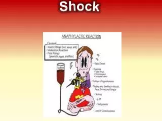
A 34 yo F presents to the ER after dining at a restaurant where shortly after eating the first few bites of her meal, became anxious, diaphoretic, began wheezing, noted diffuse pruritic rash, nausea, and a sensation of her “throat closing off”. She is currently hypotensive, tachycardic and ill appearing. Types of Shock Hypovolemic Septic Cardiogenic Anaphylactic Neurogenic Obstructive What Type of Shock is This? Anaphylactic
Anaphylactic Shock- Diagnosis • Clinical diagnosis • Defined by airway compromise, hypotension, or involvement of cutaneous, respiratory, or GI systems • Look for exposure to drug, food, or insect • Labs have no role
Anaphylactic Shock- Treatment • ABC’s • Angioedema and respiratory compromise require immediate intubation • IV line , cardiac monitor, pulse oximetry • IVFs, oxygen • Epinephrine • Second line • Corticosteriods • H1 and H2 blockers
A 41 yo M presents to the ER after an RTA complaining of decreased sensation below his waist and is now hypotensive, bradycardic, with warm extremities Types of Shock Hypovolemic Septic Cardiogenic Anaphylactic Neurogenic Obstructive What Type of Shock is This? Neurogenic
Neurogenic Shock- Treatment • A,B,Cs • Remember c-spine precautions • Fluid resuscitation • Keep MAP at 85-90 mm Hg • If crystalloid is insufficient use vasopressors • Search for other causes of hypotension • For bradycardia • Atropine • Pacemaker
A 24 yo M presents to the ED after an RTA c/o chest pain and difficulty breathing. On Physical examination, you note the patient to be tachycardic, hypotensive, hypoxic, and with decreased breath sounds on left Types of Shock Hypovolemic Septic Cardiogenic Anaphylactic Neurogenic Obstructive What Type of Shock is This? Obstructive