Download

1 / 40
410 likes | 1.32k Vues
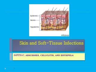
SOFT TISSUE INFECTIONS CH 152. Cathy Bulgrin DO Patty Dwyer DO. Necrotizing Soft Tissue Infections. Differentiated by primarily by depth Polymicrobial, mixed aerobic and anaerobic Early recognition and aggressive treatment important due to rapid progression and high mortality.

E N D
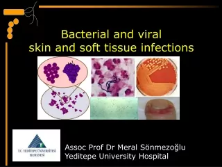
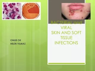
SOFT TISSUE INFECTIONSCH 152 Cathy Bulgrin DO Patty Dwyer DO
Necrotizing Soft Tissue Infections • Differentiated by primarily by depth • Polymicrobial, mixed aerobic and anaerobic • Early recognition and aggressive treatment important due to rapid progression and high mortality
Gas Gangrene (Clostridium Myonecrosis) • Rapidly progressive and limb/life threatening • Spore-forming Clostridial sp • Deepest necrotizing soft tissue infection • Hallmarks are severe myonecrosis with gas production and sepsis
Gas Gangrene (Clostridial Myonecrosis)Epidemiology • 1,000 cases per year in US • Ubiquitous organisms • 7 species, C.perfringes 80-95% • Gram +, spore forming anaerobic bacilli • Found in soil, GI and female GU
Gas Gangrene (Clostridial Myonecrosis)Pathophysiology • Produce over ten exotoxins • Exotoxin(α toxin) direct cardiodepressant, secondarily effects tissue breakdown • Mechanisms of infection are direct innoculation (open wound), hematogenous spread
Gas Gangrene (Clostidial Myonecrosis)Clinical Features • Incubation < 3 days • Pain out of proportion to physical findings • “heaviness” of affected part • Brawny edema and crepitance (later findings) • Bronze/brownish with malodorous serosanguineous d/c, bullae may be present • Low grade fever, tachycardia • Confusion, irritability or sensorium changes
Gas Gangrene (Clostidial Myonecrosis)Clinical Features Cont • Labs: metabolic acidosis, leukocytosis, anemia, thrombocytopenia, coagulopathy, myoglobinuria, myoglobinemia, liver/kidney dysfunction • GS: pleomorphic gram-positive bacilli with or without spores • Radiologic studies may demonstrate gas
Gas Gangrene (Clostidial Myonecrosis)Treatment • Resuscitation: crystalloid, plasma, packed cells • Antibiotics: PCN G (24 m units IV divided) plus clindamycin (900 mg IV q8h), ceftriaxone and erythromycin alternatives Mixed infections require aminoglycosides, PCNase resistant PCN’s or vancomycin. Tetanus as indicated. • Surgery: debridement is mainstay 4) Hyberbaric oxygen (HBO): after debridement
Gas Gangrene (Nonclostridial Myonecrosis) • Mixed infections involving aerobic and anaerobic • Presentation, eval and tx similar to Clostridial sp • Pain not as pronounced, delay in presentation • Broad-spectrum coverage: unasyn, zosyn, timentin, meropenem or imipenem • Vanc, FQ and clindamycin in PCN allergic • Early debridement and HBO
Streptococcal Myositis • Rare form of invasive group A Streptococcus • No gas production, very virulent • High rate of bacteremia and subsequent TSS • Mortality 80 – 100 %
Necrotizing FasciitisEpidemiology • 27/10,000 hospital admits • Necrosis involving SQ and fascia (no muscle) • “flesh-eating bacteria” • LE, UE, perineum, trunk, head, neck and buttocks in decreasing order of incidence • Overall mortality 25 – 50%
Necrotizing FasciitisPathophysiology • Mixed-organism most common • If single organism, typically group A strep • Symbiotic relationship between bacteria • Insults such as IV injections, surgical incisions, abscess, insect bites and ulcers • DM, PVD, smoking, IV drugs are risk factors
Necrotizing FasciitisClinical Features • Pain out of proportion to physical exam • Skin erythematous and edematous • Discoloration, vesicles, and crepitus late • Low grade fever, tachycardia are common • Early, sensorium typically clear
Necrotizing FasciitisDiagnosis • CBC with diff, chemistry with LFT’s, ABG, coags, serum lactate, blood cultures, tissue cultures • Tissue biopsy down to deep fascial plane • The “finger test”: local anesthesia, 2-cm incision into suspected area (deep fascial plane), lack of bleeding and foul smelling cloudy fluid suggestive, place finger in incision, just superior to deep fascia and push forward, if finger dissects ST away from fascia without difficulty
Necrotizing FasciitisTreatment • Aggressive fluid and resuscitation • Avoidance of vasopressors • Antibiotics similar to nonclostridial myonecrosis: empiric imipenem, meropemen or vancomycin, in PCN allergic clindamycin and FQ • Surgical debridement mainstay • HBO
Necrotizing FasciitisGroup A Streptococcus (GAS) • Presentation, eval and treatment similar to polymicrobial • Concomitant varicella infection especially in children, NSAIDs increase risk • Usually no gas formation in soft tissue • More rapid progression to bacteremia and TSS • Broad spectrum antibiotics • Clindamycin synergistic effect with PCN
Necrotizing Cellulitis • Limited to skin and SQ, polymicrobial • C. perfringes most common • Pain and erythema at infection site • Ecchymotic or frankly necrotic center • Systemic symptoms may be mild or absent • Debridement and broad spectrum antibiotics
Cellulitis • Pain, induration, warmth and erythema • Mostly staph or strep in adults, H. influenza in children • In patients with underlying disease, blood cultures and leukocytes indicated • May require doppler to differentiate DVT
Cellulitis Treatment • Dicloxicillin, macrolide, azithromycin, clarithromycin, amox-clavulanate for healthy outpatient • If head/neck, admission for IV recommended • IV meds include cefazolin, nafcillin, or oxacillin • DM, ceftriaxone, imipenem or meropenem • Ancef and probenacid, effecacious as rocephin daily • Evidence of bacteremia or underlying disease, admission to hospital
Erysipelas • Superficial cellulits involving lymphatics • Primarily GAS • Abrupt onset, high fevers, chills, malaise • Erythema with burning sensation, continues red, shiny hot plaque forms • Toxic striations and local lymphadenopathy • PenG in non DM • Nafcillin, oxacillin, rocephin, augmentin in DM • Admission to hospital
Cutaneous Abscesses Tender, swollen, erythematous, fluctuant nodule Scalp, trunk and extremity staph Oral and nasal mucosa strep Intertriginous/perineal gram negative aerobes (E.coli, P. mirabilis, Klebsiella sp) Axilla P. mirabilis Perirectal/genital anaerobic and aerobic (bacteroides sp)
Cutaneous Abscesses, Cont Foreign bodies S. aureus Cat bites Pasturella multicida, S. aureus, S. viridans, Eikenella corrodens Human bites P. multicida, Bacteroides fragilis and Corynebacterium jeikeium, staph and strep IV drugs mixed with anaerobic prevailing
Diagnosis of Cutaneous Abscess • No need for further eval if simple, healthy pt • Fever, tachycardia suggests systemic • DM, alcoholism, immunocompromised • CBC and ESR to evaluate for systemic • Immunocompromised demonstrating systemic infections need blood cultures • Foreign bodies need plain films +/- US
Treatment of Cutaneous Abscesses • Consent obtained, complications explained • If pus, I & D • If no pus, warm compresses and antibiotics • Regional or field blocks, some may require systemic sedation or OR • Area prepped and draped in sterile fashion • No. 11 or 15 scalpel, hemostats for loculated areas, irrigated and packed with gauze tape
Treatment of Cutaneous Abscesses, Cont • Warm compresses and soaking TID • F/U 2-3 days, replace packing if needed • Use of antibiotics controversial • DM, alcoholics, immunocompromised, pt with systemic symptoms should receive antibiotics • Involving hands or face, more aggressive • Antibiotic aimed at pathogen/location • Duration 5-7 days • Be aware of bacterial endocarditis
Hidradenitis Suppurativa • Recurrent chronic infection of follicle within apocrine gland • Occur in axilla, groin and perianal regions • Higher in women and AA • Usually staph, can be strep • I & D, surgeon referral, antibiotics if areas of cellulitis or systemic symptoms
Infected Sebaceous Cyst • Erythematous, tender nodule, often fluctuant • I & D • Capsule must be removed at follow up visit
Pilonidal Abscess • Superior gluteal fold • Staph most common • I & D, removing all hair and debris, packed with iodoform gauze, repacking 2 -3 days • Surgical referral
Staphylococcal Soft Tissue Abscesses Folliculitis = inflammation of hair follicle Tx: warm soaks Furuncle (boil) = abscess of hair follicle Tx: warm compresses to promote drainage Carbuncle = coalescing furuncles, large infection Tx: surgical excision
Sporotrichosis • Mycotic infection cause by Sporothrix schenkii • Commonly found on plants, vegetation and soil • Incubation period 3 weeks, 3 types of reactions, painless nodule or papule, then SQ nodules • Fungal culture, tissue biopsy diagnostic • Increased WBC, eosinophils, ESR • Itraconazole 100 - 200mg QD for 3 – 6 months
Gas Gangrene may present as: A. Pain out of proportion and heaviness B. Crepitance C. Bronze/brownish edema with malodorous discharge D. Confusion E. All of the above • Treatment of necrotizing fasciitis includes all the following except: A. Aggressive fluids and resuscitation B. Empiric antibiotics C. Vasopressors D. Surgical debridement E. HBO
3. T/F In Group A Strep Necrotizing Fasciitis, clindamycin has a synergistic effect with PCN • T/F Cutaneous abscess of scalp, trunk and extremity are usually Strep sp. • T/F Sporotrichosis incubation 3 days, treatment 3 weeks 1. E, 2. C, 3. T, 4. F staph, 5. F 3 weeks, 3 – 6 months