Download
1 / 30
300 likes | 401 Vues
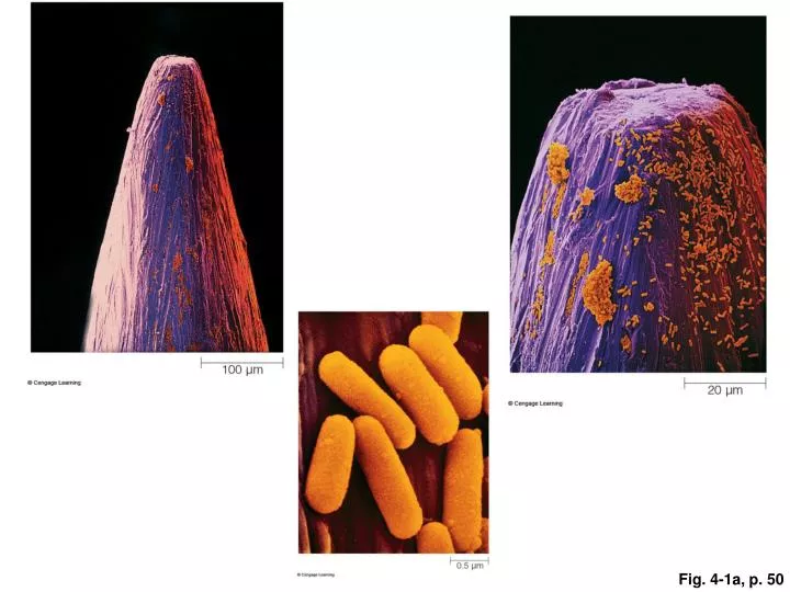
Fig. 4-1a, p. 50. Fig. 4-2, p. 51. cytoplasm. DNA. plasma membrane. a Bacterial cell (prokaryotic). Fig. 4-3, p. 52. cytoplasm. DNA in nucleus. plasma membrane. b Plant cell (eukaryotic). Fig. 4-3, p. 52. cytoplasm. DNA in nucleus. plasma membrane. c Animal cell (eukaryotic).

E N D
cytoplasm DNA plasma membrane a Bacterial cell (prokaryotic) Fig. 4-3, p. 52
cytoplasm DNA in nucleus plasma membrane b Plant cell (eukaryotic) Fig. 4-3, p. 52
cytoplasm DNA in nucleus plasma membrane c Animal cell (eukaryotic) Fig. 4-3, p. 52
extracellular environment one layer of lipids one layer of lipids membrane protein cytoplasm Fig. 4-4, p. 53
An ATP synthase, which makes ATP when H+ crosses a membrane through its interior. A calcium pump moves calcium ions across the membrane; requires ATP energy. A glucose transporter allows glucose to cross the membrane through a channel in its interior. EXTRACELLULAR FLUID LIPID BILAYER B cell receptor. It binds to bacteria, other foreign agents. Recognition protein that identifies a cell as belonging to one’s own body. CYTOPLASM phospholipid protein filaments of the cytoskeleton Fig. 4-9, p. 57
cytoplasm, with ribosomes bacterial flagellum Most prokaryotic cells have a cell wall outside the plasma membrane, and many have a thick, jellylike capsule around the wall. DNA in nucleoid plasma membrane cell wall pilus capsule Fig. 4-11, p. 58
plasma membrane nucleus mitochondria Fig. 4-14, p. 60
cell wall plasma membrane central vacuole nucleus chloroplast Fig. 4-14, p. 60
nuclear envelope chromatin nucleolus cytoplasm Fig. 4-15, p. 61
nucleus pore across the nuclear envelope nucleoplasm nucleolus chromatin nuclear envelope’s outer lipid bilayer merging with an ER membrane Fig. 4-15, p. 61
nuclear pore nuclear envelope (two lipid bilayers) cytoplasm Fig. 4-15, p. 61
chromatin pore cytoplasm nucleolus nuclear envelope (two lipid bilayers) ribosome vesicle the cell nucleus rough ER Fig. 4-16, p. 62
smooth ER channel, cross-section budding vesicle plasma membrane smooth ER Golgi body Fig. 4-16, p. 62
outer membrane outer compartment inner compartment inner membrane Fig. 4-17, p. 63
two outer membranes stroma thylakoids (inner membrane system folded into flattened disks) Fig. 4-18, p. 63
CELL WALL CHLOROPLAST CENTRAL VACUOLE NUCLEUS nuclear envelope nucleolus DNA in nucleoplasm CYTOSKELETON microtubules microfilaments intermediate filaments (not shown) RIBOSOMES ROUGH ER MITOCHONDRION SMOOTH ER PLASMODESMA GOLGI BODY PLASMA MEMBRANE LYSOSOME-LIKE VESICLE a Typical plant cell components. Fig. 4-19, p.65
NUCLEUS nuclear envelope nucleolus DNA in nucleoplasm CYTOSKELETON microtubules microfilaments intermediate filaments RIBOSOMES ROUGH ER MITOCHONDRION SMOOTH ER CENTRIOLES GOLGI BODY LYSOSOME PLASMA MEMBRANE b Typical animal cell components. Fig. 4-19, p. 64
tubulin subunit 25 nm Fig. 4-24, p. 68
actin subunit 5–7 nm Fig. 4-24, p. 68
one polypeptide chain 8–12 nm Fig. 4-24, p. 68
pair of microtubules in a central sheath protein spokes plasma membrane pair of microtubules dynein arms dynein arms basal body Fig. 4-27, p. 69