Download
1 / 47
530 likes | 1.29k Vues
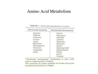
Amino Acid Structure. Dr. Azin Nowrouzi. Peptides and proteins. Polymers of amino acids. Two amino acids join covalently through a peptide bond. Another name for peptide bond is amide bond or linkage. Peptide bond formation. There are only 3 known ways to make a peptide bond

E N D
Amino Acid Structure Dr. Azin Nowrouzi
Peptides and proteins • Polymers of amino acids. • Two amino acids join covalently through a peptide bond. • Another name for peptide bond is amide bond or linkage.
Peptide bond formation • There are only 3 known ways to make a peptide bond • Chemical abiotic synthesis in the laboratory. • Genetic engineering cloning mechanisms. • Biologically in cells. • Peptide bonds in proteins are quite stable, with an average half-life (t1/2) of about 7 years under most intracellular conditions. water is removed This is a condensation reaction.
Dipeptide: 2 amino acids (aas) joined by 1 peptide bond. • Tripeptide: 3 aas joined by 2 peptide bonds. • Tetrapeptide: 4 aas joined together by 3 peptide bonds. • Peptapeptide and so forth…. • Oligopeptide: Few aas joined together by peptide bonds. • Polypeptide: Many aas joined. Molecular weights generally below 10000. • Proteins: Many aas joined. Generally have high molecular weights.
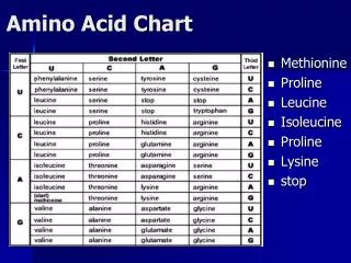
Nomenclature • The pentapeptide serylglycyltyrosylalanylleucine, or Ser–Gly–Tyr–Ala–Leu. • Peptides are named beginning with the amino terminal residue, which by convention is placed at the left. • The peptide bonds are shaded in yellow; the R groups are in red.
Ionization behavior of peptides amino-terminal (C-terminal) residue Alanylglutamylglycyllysine carboxyl-terminal (C-terminal) residue Like free amino acids, peptides have characteristic titration curves and a characteristic isoelectric pH (pI) at which they do not move in an electric field.
Length of polypeptide chains • Lengths vary considerably.
How to calculate the number of amino acids in a protein • We can calculate the approximate number of amino • acid residues in a simple protein containing no other chemical constituents by dividing its molecular weight by 110. • Although the average molecular weight of the 20 common amino acids is about 138, the smaller amino acids predominate in most proteins. • If we take into account the proportions in which the various amino acids occur in proteins, the average molecular weight of protein amino acids is nearer to 128. • Because a molecule of water (Mr 18) is removed to create each peptide bond, the average molecular weight of an amino acid residue in a protein is about 128 -18 = 110.
Polypeptides Have CharacteristicAmino Acid Compositions • The 20 common amino acids almost never occur in equal amounts in a protein. • Some amino acids may occur only once or not at all in a given type of protein; others may occur in large numbers.
Levels of Protein Structure • -carbons of adjacent amino acids are • separated by three covalent bonds • C-C-N-C • Tetrahedral angles: • N-C bond is labeled (phi). • C-C bond is labeled (psi). Rigidity of the peptide bond Primary structure Secondary structures Tertiary structure Quaternary structure • Repetitive structures • -Helix • -Sheet • parallel • antiparallel • Non-repetitive structures • -turn Amino acid sequence
Primary structure • A description of all covalent bonds (mainly peptide and disulfide bonds) linking amino acid residues in a polypeptide chain. • The linear sequence of amino acids within a peptide • Written from NC, • either in three-letter code, • or, more often, in one-letter code. • Example: Glu-Gly-Ala-Lys or EGAK
Determination of amino acid composition of a polypeptide • Primary structure determination • Pure sample must be used. • Acid hydrolysis: strong acid, 110 C, 24hr. • Chromatography: Cation exchange, each amino acid exits the column at a specific pH and ionic strength. • Quantitative analysis by heating with ninhydrin • This analysis is done by amino acid analyzer.
Sequencing the peptide from N-terminal • Phenylisothiocyanate (Edman’s reagent) is used to label the amino-terminal residue under mildly alkaline conditions. • The resulting phenylthiohydantoin (PTH) derivative causes an instability in the N-terminal peptide bond. • This bond can be hydrolyzed without cleaving the other peptide bonds. • This procedure can be applied repeatedly to the shortened peptide. • Good for about 100 aa. • Has been automated (sequenator)
Cleavage of polypeptide chain into smaller fragments • When more than 100 aa are present in a polypeptide chain. • By using more than one cleaving agent on separate samples of the purified polypeptide, overlapping fragments are generated. • Enzyme cleavage, such as trypsin and other digestive enzymes • Chemical cleavage (cyanogen bromide) • Overlapping peptides
Specific cleavage of polypeptides • Proteins larger than 50 aa are first hydrolyzed into shorter peptides. • Chemical or enzymatic methods hydrolyze proteins at specific sites. • Peptides are separated by chromatography • Peptides generated by 2 or more cleavage methods are each sequenced separately. • Sequences of individual peptides are overlapped together to deduce the entire protein sequence
a-Helix • H-bonds are inside the chain
Description of -helix • The polypeptide backbone is tightly wound around an imaginary axis drawn longitudinally through the middle of the helix. • The R groups of the amino acid residues protrude outward from the helical backbone. • Each helical turn includes 3.6 amino acid residues. • About one-fourth of all amino acid residues in polypeptides are found in -helices.
Stabilization of -helix • The structure is stabilized by a hydrogen bond between the hydrogen atom attached to the electronegative nitrogen atom of a peptide linkage (amino acid n) and the electronegative carbonyl oxygen atom of the fourth amino acid (amino acid n+4) on the amino-terminal side of that peptide bond. • Each successive turn of the -helix is held to adjacent turns by three to four hydrogen bonds. • All the hydrogen bonds combined give the entire helical structure considerable stability. • Naturally occurring L-amino acids can form either right- or left-handed helices, but extended left-handed helices have not been observed in proteins.
Factors affecting stability • Electrostatic repulsion (or attraction) between successive amino acid residues with charged R groups. • Bulkiness of adjacent R groups. • The interactions between R groups spaced three (or four) residues apart. • Presence of Pro or Gly residues. • Proline is only rarely found within an helix • In proline, the nitrogen atom is part of a rigid ring and rotation about the N-Cbond is not possible. Thus, a Pro residue introduces a destabilizing kink in an helix. • In addition, the nitrogen atom of a Pro residue in peptide linkage has no substituent hydrogen to participate in hydrogen bonds with other residues. • Glycine occurs infrequently in helices for a different reason • It has more conformational flexibility than the other amino acid residues. • Polymers of glycine tend to take up coiled structures quite different from an -helix. • Interaction between amino acid residues at the ends of the helical segment and the electric dipole inherent to the helix.
Helix dipole • A net dipole extends along the helix that increases with helix length. • Negatively charged amino acids are often found near the amino terminus of the helical segment, where they have a stabilizing interaction with the positive charge of the helix dipole. • A positively charged amino acid at the aminoterminal end is destabilizing.
Secondary structure: Fully extended chains can form -sheets
Non-repetitive secondary structures • Turns • Connections • Loops • Coils or random coils • These are well ordered but non repeating configurations.
turns • type I turns occur more than twice as frequently as type II. • Type II turns always have Gly as the third residue.
Tertiary structure • Amino acids that are far apart in the polypeptide sequence and that reside in different types of secondary structure may interact within the completely folded structure of a protein. • The location of bends (including turns) in the polypeptide chain and the direction and angle of these bends are determined by the number and location of specific bend-producing residues, such as Pro, Thr, Ser, and Gly. • Interacting segments of polypeptide chains are held in their characteristic tertiary positions by different kinds of weak bonding interactions (and sometimes by covalent bonds such as disulfide cross-links) between the segments. Leptin
Stabilization of tertiary Structure • overall three-dimensional arrangement of all atoms in a protein is referred to as the protein’s tertiary structure. • Stabilized primarily through weak bonds.
Three-dimensional structures of some small proteins • PDB; www.rcsb.org/pdb Lysozyme PDB ID 3LYM RibonucleasePDB ID 3RN3 Myoglobin PDB ID 1MBO Cytochrome c PDB ID 1CCR
Interactions stabilizing tertiary structure • Specific overall shape of a protein • Cross links between R groups of amino acids in chain
Domains • When molecular weight is larger than 20000. • The ratio of surface area to volume is small. • A protein with multiple domains may appear to have a distinct globular lobe for each domain.
Example • Crystal structure of the heterodimeric enzyme Rab Geranylgeranly Transferase. • It is a dimer of a alpha (blue, red, yellow) and a beta subunit (orange). • The alpha subunit is a multi domain protein.
Quaternary structure • Protein Quaternary Structures Range from Simple Dimers to Large Complexes: • A multisubunit protein is also referred to as a multimer. • Multimeric proteins can have from two to hundreds of subunits. • A multimer with just a few subunits is often called an oligomer. • The repeating structural unit in such a multimeric protein, whether it is a single subunit or a group of subunits, is called a protomer. • The first oligomeric protein for which the three dimensional structure was determined was hemoglobin (Mr 64,500), which contains four polypeptide chains.
Hemogloin A tetrameric protein two a-chains (141 AA) two b-chains (146 AA) four heme cofactors, one in each chain The a and b chains are homologous to myoglobin. Oxygen binds to heme in hemoglobin with same structure as in Mb but cooperatively: as one O2 is bound, it becomes easier for the next to bind.
Simple and conjugated proteins • Some proteins contain chemical groups other than amino acids.
Spectroscopy of amino acids • Aromatic amino-acids are strong chomophores in the far-uv. • Only the aromatic amino acids absorb light in the UV region
Ninhydrin-detection of amino acids • Complete hydrolysis for 24 hr at 110 oC in 6 M HCl. • Amino acids can be detected on the chromatogram by using ninhydrin. A solution of ninhydrin is sprayed onto the paper and heated. The amino acids show up as purple spots(proline appears yellow).
Paper chromatograms 2D chromatogram eluting with a different solvent mixture in each direction.
Electrophoresis • Electrophoresis is a technique that uses the net charge of peptides (amino acids) as a basis for separation. • A potential difference is applied across a solid material (e.g. paper for amino acid analysis) permeated by an electrolyte. • Anions migrate to the anode and cations to the cathode. The rate of diffusion is related to the size and net charge. Small highly charged proteins migrate more quickly.