Download
1 / 42
420 likes | 423 Vues
Learn about refraction, hypermetropia, astigmatism, and their management. Includes case study and explanations.

E N D
Refraction - II Dr AjaiAgrawal Additional Professor Department of Ophthalmology AIIMS, Rishikesh
Acknowledgement • Photographs in this presentation are courtesy of Kanski’sClinical Ophthalmology.
Learning Objectives At the end of the class, students shall be able to • Understand what is refraction. • Have basic knowledge of hypermetropia and astigmatism and their management.
Question • A patient with a corneal scar is carefully refracted. Best corrected visual acuity is 6/12. With a pinhole over his correction, his acuity improves to 6/6. The best explanation for this is • a. spherical aberration. • b. myopic astigmatism. • c. cataract. • d. irregular astigmatism.
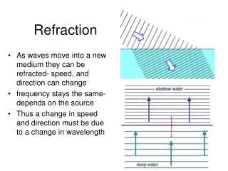
What is Refraction • When rays of light traveling through air enter a denser transparent medium, the speed of the light is reduced and the light rays proceed at a different angle, i.e., they are refracted. • Except when the rays are normal Refraction in Ophthalmology • Methods for evaluating the optical and refractive state of the eye
Emmetropia • Parallel light rays, from an object more than 6 m away, are focused at the plane of the retina when accomodation is at rest. • Clear image of a distant object formed without any internal adjustment of the optics of the eye. • Absence of emmetropia = Ametropia
Refractive errors Anomalies of the optical state of the eye Myopia Hypermetropia Astigmatism
Hypermetropia • Refractive or Diopteric state of eye wherein incident parallel rays of light coming from infinity are focused behind the retina with accommodation being at rest.
Near images can be blurred unless there is sufficient accommodation, as in a child. • They have blurred images for distant objects also • Most children are born about +3 D hyperopic, but this usually resolves by age 12 years.
Types | | | | | | Axial Curvature Index Positional Absence of lens • Axial is the commonest form. • In this condition the total refractive power of eye is normal but there is axial shortening of eye ball.
Each millimeter of shortening represents approximately 3D of refractive change and thus a hypermetropia of over 6D is uncommon. • Physiological: Infant, child. • Pathological: Orbital tumour, or inflammatory mass may indent the posterior pole of the eye and flatten it
Curvature Hypermetropia : When the radius of curvature of any of the refracting surfaces is increased, • congenitally (cornea plana) or as a result of trauma • Increase of 1 mm produces a hypermetropia of 6 D.
Index Hypermetropia : Usually manifests itself as a decrease in the effective refractivity of the lens and is responsible for the hypermetropia which occurs physiologically in old age and pathologically in diabetes.
Positional Hypermetropia : Posterior placed lens also produced hypermetropia whether it occurs as a congenital anomaly or as a result of trauma and disease. • Aphakia: Surgical, posterior dislocation of lens
Clinical Types: • Simple Hypermetropia : Commonest form. • It results from normal biological variations in the development of eye e.g., axial and curvatural. • Pathological Hypermetropia : Either congenital or acquired conditions of eyeball which are outside the normal biological variation of development • Example: index , positional (Aphakia). • Functional Hypermetropia : Results from paralysis of accommodation as seen in patients with third nerve palsy.
Components of hypermetropia Total hypermetropia = Latent+manifest(facultative + absolute) Accommodation in Hypermetropia • Contraction of ciliary muscle in the act of accommodation increases the refractive power of the lens so that it corrects a certain amount of hypermetropia.
Normally there is an appreciable amount corrected by contraction involved in physiological tone of ciliary muscle. • Consequently the full degree of hypermetropia is revealed only when this muscle is paralysed by the use of a drug such as atropine. • This is called latent hypermetropia, normally 1D.
Manifest Hypermetropia consists of: • Facultative Hypermetropia: Corrected by effort of accomodation • Absolute Hypermetropia: Cannot be overcome by effort of accomodation • As tone of ciliary muscle decreases with age, some latent hypermetropia becomes manifest • As range of accomodation reduces with age, more facultative hypermetropia becomes absolute, all of it after age 60.
Symptoms • Vary with degree of hypermetropia and accomodative effort • Blurred vision: near>distant • Accomodativeasthenopia • Convergent squint due to continuous effort of accomodation, excess of convergence leads to dissociation of muscle balance • Early onset of presbyopia
Signs • Small eyeball • Smaller cornea • Shallow anterior chamber predisposes to angle closure glaucoma since size of lens is normal • Apparent divergent squint
Retina : Has peculiar sheen : a reflex effect so called “shot silk retina” on ophthalmoscopy. • Optic disc : Characteristic appearance which may resemble optic neuritis (Pseudopapillitis).
Treatment • In young children below the age of 6-7 years: some degree of hypermetropia is physiological and a correction need be given only if the error is high or if strabismus is present. • In those between 6 and 16 years: smaller error may require correction.
Refractive correction is required • in middle aged patients • in high hypermetropia • and if patient is symptomatic • Optical: • Glasses • Contact lens
Convex lenses prescribed after full cycloplegic refraction, particularly in children • Child with convergent squint may need “full atropine correction” • Contact lens power is a little more than spectacle power
Surgical Options • Conductive keratoplasty. • Non contact Holmium YAG laser thermokeratoplastyfor lower hypermetropia (+1D – 2.5 D). • Phakic Intraocular lens (+6D – +10 D)
Astigmatism • Astigmatism is a type of refractive error wherein the refraction varies in different meridia. • Consequently rays of light entering the eye cannot converge to a point focus, but form focal lines. Astigmatism | | | Regular Irregular
Astigmatism • Light rays passing through a steep meridian are deflected more than those passing through a flatter meridian.
1. Corneal Astigmatism e.g. keratoconus 2. Lenticular Astigmatism (i) Curvatural – e.g. lenticonus (ii) Positional – subluxation (iii) Index – cataract 3. Retinal astigmatism – due to oblique placement of macula.
Types of Regular Astigmatism • With the rule astigmatism : The two principal meridia are placed at right angles to one another but the vertical meridian is more curved than horizontal meridian • (most common type) • Against the rule astigmatism : Horizontal meridian is more curved than the vertical meridian. • 3. Oblique astigmatism : Is a type of regular astigmatism where the two principal meridia are not horizontal and vertical , though they are at right angles to one another (45 and 135 deg)
Oblique astigmatism : (i) Symmetrical : Cylindrical lens required at same axis in both eyes. (ii) Complementary : Cylindrical lens required at 30o in one eye and at 150o in the other eye. 4. Bi-oblique astigmatism : In this type of regular astigmatism the two principal meridia are not at right angles to each other, one eye at 30o and other at 100o.
Optics of regular astigmatism : In regular astigmatism the parallel rays of light are not focused on a point but form two focal lines – Sturm’s conoid
Refractive types of Regular astigmatism • Depending upon the position of two focal lines in relation to retina, regular astigmatism is further classified • Simple : Where one focus falls upon retina, the other focus may fall in front of or behind, so that one meridian is emmetropicthe other is either hypermetropic or myopic.
Compound : Where neither of two foci lie upon retina but both are placed in front or behind it. The state of the refraction is then entirely hypermetropic or entirely myopic. The former is known as compound hypermetropic, the latter as compound myopic astigmatism.
3. Mixed : Where one focus is in front of and other behind retina so that the refraction is hypermetropic in one direction and myopic in the other.
Irregular Astigmatism : Refraction in different meridia are irregular. • Etiological types: • Curvaturalirregular astigmatism: irregular healing of cornea after trauma and inflammation (particularly ulceration & keratoconus) • Index irregular astigmatism : incipient cataract
Symptoms • Defective vision • Blurring of objects • Asthenopic symptoms - eyeache and headache • Running of lines
Treatment • Optical – Spectacles with cylindrical lenses, Contact lens (Toriccontact lenses with prism ballast) • Surgical • Astigmatic keratotomy: LimbalRelaxing Incision, arcuatekeratectomy, removal of sutures • Photo-astigmatic refractive keratotomy (PARK) • Laser: Excimer laser: LASIK or Femtosecond laser
Guidelines for Optical treatment • If the patient does not complain of asthenopic symptoms small astigmatic errors (0.5 D or less) generally do not require correction • If asthenopic symptoms are present , error should be corrected by cylindrical lenses. • Undercorrect the error initially • At a later date, full correction may be worn comfortably.
Question • In a patient with astigmatism, all of the following are true of myopia and hyperopia except • a. In simple myopic astigmatism, one focal line lies in front of the retina and the other is on the retina. • b. In compound myopic astigmatism, both focal lines lie in front of the retina. • c. In simple hyperopic astigmatism, both focal lines lie behind the retina. • d. In mixed astigmatism, one focal line lies in front of the retina and one lies behind the retina.