Download
1 / 118
1.48k likes | 2.62k Vues
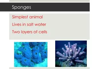
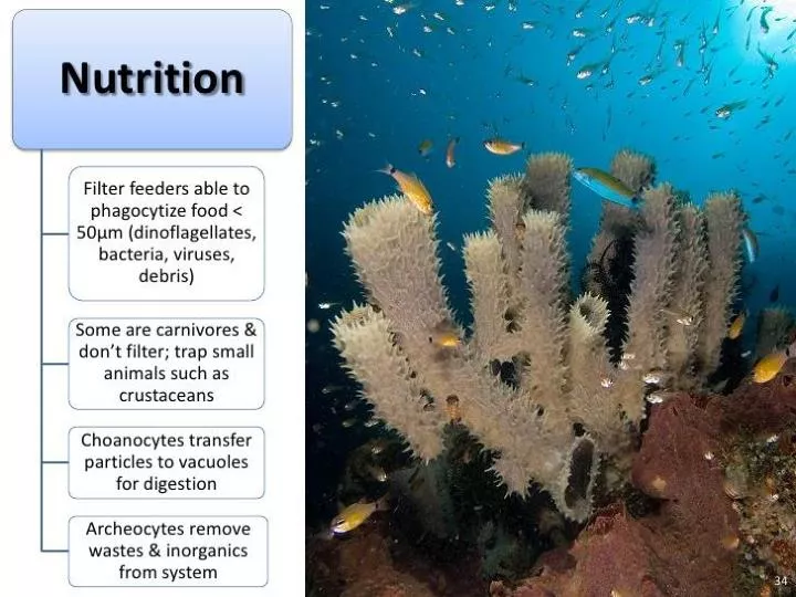
Feeding in Sponges. Particles larger than 50 micrometers cannot enter the ostia and pinacocytes consume them by phagocytosis (engulfing and internal digestion). Particles from 0.5 to 50 micrometres are trapped in the ostia, which taper from the outer to inner ends.

E N D
Feeding in Sponges • Particles larger than 50 micrometers cannot enter the ostia and pinacocytes consume them by phagocytosis (engulfing and internal digestion). • Particles from 0.5 to 50 micrometres are trapped in the ostia, which taper from the outer to inner ends. • These particles are consumed by pinacocytes or by archaeocytes which partially extrude themselves through the walls of the ostia.
Feeding • Bacteria-sized particles, below 0.5 micrometers, pass through the ostia and are caught and consumed by choanocytes. • Since the smallest particles are by far the most common, choanocytes typically capture 80% of a sponge's food supply. • Archaeocytes transport food packaged in vesicles from cells that directly digest food to those that do not. • At least one species of sponge has internal fibers that function as tracks for use by nutrient-carrying archaeocytes, and these tracks also move inert objects.
FEEDING The series of 4 drawings shows one food-transfer option. After a bacterium is phagocytosed, the choanocyte resorbs its collar and crawls off into the mesohyl to distribute the digested food products,possibly to an amoebocyte
Pseudopodia (lamellipodia) wrapping around larger beads (1um) at the choano-cyte surfaces
Carnivorous Sponges • Some species in the family Cladorhizidae capture and digest whole animals. • Capture small crustaceans with their spicules, which 'hook onto' and attach to crustacean exoskeletons that they touch. • Sponge cells then migrate around the prey, and digestion takes place extracellularly.
Cladorhizids- carnivorous sponges • Class Demospongia, Family Cladorhizidae • Discovered in 1995 • Abyssal and troglobytic • Spicules act as hooks to snare small invertebrates Asbestopluma bihamatifera
Chondrocladia lyra Chondrocladia gigantea
Chondrocladia lampadiglobus Asbestopluma hypogea
Reproduction and Development • Asexual Reproduction • Fragmentation • See observations from lab exercise • Buds • Gemmules • Overwintering bodies • Thick covering of spongin
Micropyle Spicule Mass of archaeocytes Sponge Gemmule Overwintering protective body of a sponge
Regeneration • When sponge is pushed through fine mesh strainer • Cells will reaggregate and begin dividing to reconstitute the sponge • Cells from different species will not aggregate • New sponges can grow from broken fragments • See lab exercise
Reproduction and Development • Sexual Reproduction • Highly variable • Most sponges are hermaphrodites • Both male and female gametes are produced by the same individual • Eggs from choanocytes and archaeocytes • Sperm from choanocytes • BUT most produce eggs at one time and sperm at a different time • What is the significance of this?
Reproduction and Development • Sexual Reproduction • Fertilization is external in some • Takes place in the water • “ovipary” • Planktonic larvae
Reproduction and Development • Sexual Reproduction • Fertilization is internal in some • Choanocytes trap sperm • De-differentiate to amoebocytes = carrier cell • Transport sperm to eggs in the mesohyle • Larval development • Some species retain the fertilized eggs • Early development is internal • Embryos are released as swimming larvae • Leave through excurrent pores or rupture parent • viviparous
Cross section, of Grantia showing larval amphiblastula embedded in the body wall.
Sycon coactum - after squeezing through the choanocyte layer the larvae are carried out of the sponge in the water flow
X-section of amphiblastula larva showing flagellated cells at anterior end and an internal cavity, the blastocoel Amphiblastula larva of Sycon sp. Posterior cells later grow around to enclose the flagellated cells, which come to face inwards. The remnant of the blastocoel becomes filled with mesohyl (a proteinaceous material). An opening, the osculum, appears (at top of R-hand figure)
Reproduction and Development • Larval development • Larvae do not feed • Larvae swim for about 24 hours • Settle down • Attach to substrate • Metamorphose • Larval cells migrate and differentiate into the different cell types of the adult sponge • Free-swimming larva are dispersal stage • Make sure this is part of your 12 character chart!
Reproduction • Reproduction • Sexual • Development • Rearrangement of cells at time of settling corresponds to gastrulation stage of all other animals • In some sponges outer flagellated cells are lost at gastrulation • In the Calcaria, flagellated cells de-differentiate into mutipotent amoebocytes
3 Types of Larvae • Parenchymula Larva • Found in Demospongiae • Solid, covered with monoflagellated cells • Incubated until late in development • Repeated cleavage of the zygote takes place in the mesohyl and forms a parenchymula larva • Mass of larger internal cells surrounded by small, externally flagellated cells. • Swimming larvae enter a canal of the central cavity and are expelled with the exhalant current
Larva of Amphimedon queenslandica Parenchymula larva
3 types of Larvae • Coeloblastula Larva • Found in calcareous sponges • Ciliated, hollow blastula • Short incubation • Complex development • Micromeres • Macromeres • Intermediate stage = stomoblastula • Consumes nutrient-rich amoebocytes • Turns inside-out to form amphiblastula larva
Coeloblastula larva Plakina
The interior of the brood chamber of a sponge, Amphimedon queenslandica, showing embryos in the early phases of development.
Amphiblastula larva Amphiblastula larva of Sycon ciliatum Settled sponge larva. The flagellated cells have invaginated.