Download
1 / 1
10 likes | 119 Vues
a. b. c. NMBA, CS, and Sepsis. CS Only. d. e. f. FIGURE 3. g. h. i. Day 1. Day 5. Day 1. Day 5. FIGURE 1. A. B. Myosin mRNA expression in the EDL, soleus, diaphragm and masseter muscles of control, denervated and neuromuscular blocked mechanically ventilated rats. FIGURE 4.

E N D
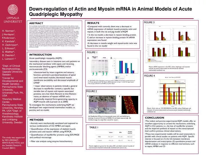
a b c NMBA, CS, and Sepsis CS Only d e f FIGURE 3 g h i Day 1 Day 5 Day 1 Day 5 FIGURE 1 A. B. Myosin mRNA expression in the EDL, soleus, diaphragm and masseter muscles of control, denervated and neuromuscular blocked mechanically ventilated rats. FIGURE 4 A) SDS-PAGE (12%) of pig gluteus maximus muscle in NMBA, CS and Sepsis treated animal and an CS only exposed animal. B) Myosin mRNA expression on days 1, 3, and 5 for pigs treated with varying combinations of NMBA, CS and/or Sepsis. FIGURE 2 A B Myosin : Actin ratio on 12% SDS-PAGE for the EDL, soleus, Diaphragm and masseter of control, denervated and neuromuscular blocked mechanically ventilated rats. (A) Myofibrillar ATPase (a-c), hematoxylin-eosin (d-f), and NADH (g-h) stainings of control, CS only and NMBA, CS and sepsis exposed pigs. (B) Confocal image of a single fiber stained for myosin. • RESULTS • Compared with controls, there was a decrease in mRNA expression of skeletal muscle proteins, actin and myosin, in both the rat and pig model of AQM • In the rat model, a decrease in myosin binding protein C and an increase in myosin binding protein H mRNA expression was found • A decrease in muscle weight and myosin:actin ratio was found in the rat model ABSTRACT Acute quadriplegic myopathy (AQM), which is characterized by symmetrical weakness and/or paralysis of spinal nerve innervated muscles and intact sensory and cognitive function, affects a large percentage of patients requiring mechanical ventilation. However, the mechanisms underlying AQM are poorly understood, but clinical studies have shown a complete or partial loss of the motor protein myosin and a block of myosin synthesis at the transcriptional level. The diagnosis of AQM is typically made after the disease has progressed to the later stages, which makes understanding the cellular and molecular mechanisms of the disease difficult. To better understand the initial mechanisms of AQM, two experimental animal models have been developed in the rat and pig. Specific interest is focused on the expression and content of the myofibrillar proteins actin and myosin using confocal microscopy and SDS-PAGE gel electrophoresis and mRNA expression using real-time PCR. In accordance with previous findings from patients with AQM, we observe a decrease in myofibrillar protein mRNA expression in spinal nerve innervated muscles such as the extensor digitorum longus, soleus and biceps femoris. Cranial innervated muscles (e.g., masseter) and those muscles passively activated (e.g., diaphragm) were less affected. Additionally, there was a decrease in myofibrillar protein expression as seen using SDS-PAGE gel electrophoresis. In conclusion, this suggests that the rodent and porcine experimental models will be very valuable in future mechanistic studies on the molecular mechanisms underlying the development of AQM. Furthermore, our data support the idea that the down-regulation of myosin and actin mRNA may play a role in the underlying mechanisms of AQM. H. Norman1, J. Nordquist1, P. Andersson1, K. Kandala2, H. Zackrisson4, L. Eriksson4, S. Walther4, B. Dworkin3, L. Larsson1,2 1Dept. of Clinical Neurophysiology, Uppsala University, Sweden 2Center for Developmental and Health Genetics , Pennsylvania State University, University Park, PA, USA 3Hershey Medical Center, Pennsylvania State University, Hershey, PA, USA 4Dept. of Anesthesiology, Karolinska Institute and Linköping University, Sweden • INTRODUCTION • Acute quadriplegic myopathy (AQM): • secondary disease seen in intensive care unit patients on the mechanical ventilator with sepsis and receiving neuromuscular blocking agents (NMBA) and/or corticosteroids (CS) • characterized by intact cognitive and sensory function, symmetric paralysis/weakness of spinal cord innervated muscles, decreased muscle membrane excitability and decreased amplitude of CMAP • major observations in patients include a general decrease in myofibrillar content, a specific but variable loss of myosin and myosin associated proteins, very low thick-filament to thin-filament ratios, an absence of myosin mRNA and a dramatically impaired force-generating capacity in AQM muscle cells (Larsson et al, 2000) • To investigate the mechanisms underlying AQM we developed two experimental mammalian models, i.e. porcine and rat model. Down-regulation of Actin and Myosin mRNA in Animal Models of Acute Quadriplegic Myopathy • CONCLUSION • The rodent and porcine experimental AQM models offer an excellent opportunity to unravel the mechanisms underlying AQM in ICU patients, i.e., the loss of myofibrillar proteins and the impaired synthesis of myosin at the transcriptional level confirm previous clinical observations. • These two experimental models will be used extensively in parallel with clinical studies on patients with AQM. Specific interest is focused on (1) the link between membrane depolarization and transcription, (2) quantitative protein and mRNA analyses in response to different interventions, such as sepsis, NMBA and CS • METHODS • Animals were mechanically ventilated and exposed to various combinations of CS, NMBA and sepsis • Quantification of the expression of skeletal muscle proteins actin and myosin mRNA using RT-PCR • Quantification of myofibrillar proteins using SDS-PAGE and confocal microscopy • Fiber size analysis using enzyme histochemistry TABLE 1 The study was supported by NIH (AR45627, AR47318, AG14731) and the Swedish Research Council (8651) Muscle weights (in grams) for the soleus, EDL and tibialis anterior muscles of neuromuscular blocked mechanically ventilated and control rats.