Download
1 / 15
150 likes | 447 Vues
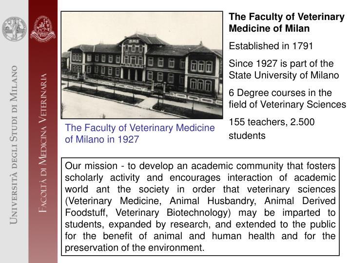
The Faculty of Veterinary Medicine of Milan Established in 1791 Since 1927 is part of the State University of Milano 6 Degree courses in the field of Veterinary Sciences 155 teachers, 2.500 students. The Faculty of Veterinary Medicine of Milano in 1927.

E N D
The Faculty of Veterinary Medicine of Milan Established in 1791 Since 1927 is part of the State University of Milano 6 Degree courses in the field of Veterinary Sciences 155 teachers, 2.500 students The Faculty of Veterinary Medicine of Milano in 1927 Our mission - to develop an academic community that fosters scholarly activity and encourages interaction of academic world ant the society in order that veterinary sciences (Veterinary Medicine, Animal Husbandry, Animal Derived Foodstuff, Veterinary Biotechnology) may be imparted to students, expanded by research, and extended to the public for the benefit of animal and human health and for the preservation of the environment.
The “Veterinary Teaching Hospital” in Lodi Since 2005 Made with the support of State University of Milano, Lombardia region, Province and Municipality of Lodi
The Faculty of Veterinary Medicine of Milano-Lodi and a project of “Veterinary TeleMedicine” • What is Veterinary TeleMedicine? • The ability to send veterinary medical data using any form of communication interface, from the patient to any other site: • Pictures or video of clinical cases • Imaging: x-ray, ultrasonography, CT, MRI or other • Pictures of gross lesions • Pictures from histological or cytological samples • Any biomedical information A little unit of Veterinary TeleRadiology is already present in Veterinary Faculty SkypeTM
Veterinary TeleMedicine as in human being • Is a point-to-point telemedicine system • Symmetric • - no distinction between nodal and referral centers • - any clinic can communicate to other clinics • - second level referral is allowed • Multi nodal, multi referral environment • Operates over a spectrum of low to high bandwidth communication channels
Veterinary TeleMedicine as in human being • Store and forward technical informations • Online video conferencing and data transfer • Electronics Medical Record (EMR) Supported • - Text, Image, Graphics, Audio, Video • Integration with different medical instrument • EEG, ECG, USG, MRI, CT SCAN, Microscope with digital camera, others • Support of medical standards
Friesian heifer, 5 months old, 150 kg (= 330 pounds) CASE HISTORY: swinging hindleg lameness RADIOLOGICAL DIAGNOSIS: luxation of the coxofemoral joint: in the blue circle is observed the acetabulum while in the red circle the femoral head
Friesian cow, 1½ years old, 450 Kg (= 990 pounds) CASE HISTORY: non-weight-bearing lameness RADIOLOGICAL DIAGNOSIS: traumatic infectious arthritis the probe (red arrows) is into the skin wound. There is osteomyelitis of the medial metacarpal-phalangeal joint and septic periostitis (yellow arrows). Multiple gas-capped fluid level indicates the presence of abscesses in the swollen soft tissues (white arrows)
M M M Friesian cow, 3 years old, 700 kg (=1540 pounds) CASE HISTORY: stop eating and chewing her cud. The “grunt test” was positive RADIOLOGICAL DIAGNOSIS: sand in the reticular cups allows good identification of the mucosal profile of the reticulum (white arrows). A standard lozenge-shape bolus magnet (M) is lying superimposed on the floor of the reticulum. Several metallic foreign bodies are adhered to the magnet, with one of them has probably penetrated the reticular wall
Clinical history: Persistent high fever , photophobia, anasarca, diarrhoea, and generalized lymphoadenopathy. Followed by a progressive thickening of the skin, exudation, fissuring, and the development of a marked alopecia. Macroscopic findings: the skin is thick, alopecic, lichenified, scaly, and focally ulcerated.
Cytologic description:presence of numerous free protozoal organism, crescent-shaped, (5-7µm), with bluish cytoplasm and small eccentric nuclei. Name of the disease, ethiology: skin: besnoitiosis (Besnoitia besnoiti) Comment:B. besnoiti is an obligate intracellular coccidian protozoa. Cattle serve as intermediate hosts of B. besnoiti .Clinical besnoitiosis in cattle is characterized by two sequential stages: an acute febrile stage and a chronic seborrheic stage. Fine needle aspiration of cutaneous lesions, May-Grünwald Giemsa stain.Numeous bradyzoites of B. besnoiti. Few lymphoid and monocytoid cells are also present.
Clinical history: fever and respiratory signs in a group of adult cattle. 30% of animals affected, 15% of mortality. Necropsy findings: sero-fibrinous exudate in the pleural space. Area of consolidation in the basal lobe of the lung with red/gray lobules and enlargement of the interstitial tissue Picture sent: lung from a cattle, basal lobe, cut surface.
Morphological diagnosis: Locally extensive, severe, subacute fibrinous pleuropneumonia Name of the disease, ethiology: Contagious bovine pleuropneumonia (CBPP), Mycoplasma mycoides subs. mycoides Suggested confirmatory laboratory tests: bacteriological culture from lung (need refrigeration), serology. Immunohistochemistry on formalin fixed samples (can be sent to our labs without refrigeration) Positive immunohistochemical staining for Mycoplasma mycoides subs. mycoides antigen (in brown)
KEY MESSAGE • New technology must be relevant for successful, but it has not to be substitute for well - trained staff in the right place at the right time Algeria: School of Veterinary Medicine in a refugee camp: Teacher shows instruments for surgery
KEY MESSAGE • Delegation of key skills to an emerging and evolving veterinary profession and animal husbandry appears essential to the objectives of veterinary telemedicine Teacher shows to the students a vaginal prolapse in a goat