Download
1 / 21
250 likes | 1.52k Vues
Reversed-Phase HPLC Analysis of Aminoglycoside Antibiotics Using Evaporative Light Scattering Detection. Laura E. Manis and Melissa J. Wilcox Alltech Associates, Inc. 2051 Waukegan Road • Deerfield, IL 60015 Phone: 1-800-ALLTECH • Web Site: www.alltechweb.com. Introduction.

E N D
Reversed-Phase HPLC Analysis ofAminoglycoside Antibiotics Using Evaporative Light Scattering Detection Laura E. Manis and Melissa J. Wilcox Alltech Associates, Inc. 2051 Waukegan Road • Deerfield, IL 60015 Phone: 1-800-ALLTECH • Web Site: www.alltechweb.com
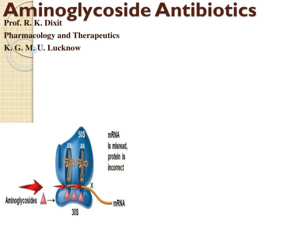
Introduction • The Evaporative Light Scattering Detector (ELSD) is becoming more common in today’s HPLC laboratories. Evaporative light scattering detection offers sensitive, universal detection of any sample that is less volatile than the mobile phase. Unlike UV and RI detectors, the ELSD’s response is independent of the sample’s optical characteristics. Therefore, any non-volatile sample, chromophoric or non-chromophoric can be detected, regardless of its functional group. Response from an ELSD is based on mass concentrations of the analyte and the amount of light scattered, which is useful in detecting unknown components and impurities. The ELSD also maintains a stable baseline over gradient runs, making it ideal for gradient applications. The ELSD answers the need for improved HPLC methods by offering universal and sensitive detection for non-volatile samples, regardless of optical activity. • Aminoglycoside (AG) antibiotics are commonly used for treating infection caused by gram-negative bacteria. A rugged HPLC method is important to scientists interested in monitoring or researching aminoglycosides. This class of antibiotics lacks a significant UV chromophore, which makes ELS detection ideal.
Other methods of detection for aminoglycosides include RI, UV, and MS detection. RI detectors are not gradient compatible, less sensitive, and susceptible to ambient temperature changes. Aminoglycoside antibiotics do not have strong chromophores, which makes detection by UV difficult and not sensitive. ELSD also offers more cost-effective analysis and less complicated operation than an MS detector. The ELSD overcomes the challenges of gradient analysis, optical response, and cost of instrumentation. • Evaporative Light Scattering Detectors use a simple three-step process that produces a signal for any non-volatile sample component. This unique detection method is the key to the ELSD’s versatility and performance. Since all particles scatter light, all non-volatile sample analytes are detected with high sensitivity and accuracy, regardless of their functional groups or optical properties. All samples are detected with nearly equivalent response factors, making concentration determination easier when authentic standards are not available.
ELSD Detection Principles – Universal, Versatile, Sensitive 1. Nebulization: Inside the nebulizer, the column effluent passes through a needle, mixes with nitrogen gas, and forms a dispersion of droplets. 2. Mobile Phase Evaporation: The droplets pass through a heated “drift tube” where the mobile phase evaporates, leaving a fine mist of dried sample particles in solvent vapor. 3. Detection: The sample particles pass through a flowcell where they are hit with a laser light beam. Light scattered by the sample particles is detected, generating an electrical signal.
Objective • This paper highlights a rugged, ELSD compatible HPLC method to analyze four common aminoglycosides (AGs): amikacin, neomycin, streptomycin, and tobramycin. The study investigates analytical column durability in the extreme low pH range and the use of buffered ion-pair reagents in retaining the antibiotics on a C18 column. A comparison of UV and ELSD shows the superiority of ELS detection for these antibiotics.
Experimental • Instrumentation: • • Alltech (Deerfield, IL) On-line Vacuum Degasser • • Alltech Model 580 Autosampler • • Alltech Model 500 ELSD (Evaporative Light Scattering Detector) • • Hitachi (Tokyo, Japan) L-6200A Intelligent Pump • • Linear (Thermo Separation Products, San Jose, CA) Model 200 UV/Vis Detector • • PE Nelson™ (Norwalk, CT) Turbochrom™ EL Datastation • Columns: • • Alltech Alltima™ C18, 5µm, 250 x 4.6mm • • Hamilton (Reno, NV) PRP™-1, 5µm, 150 x 4.6mm • • OraChrom (Woburn, MA) Styros™ 2R/XH, 3µm, 100 x 4.6mm
Reagents and Samples: • • 18.2MΩ water purified by a Millipore (Bedford, MA) Elix and Gradient system • • HPLC grade solvents were purchased from Burdick and Jackson (Muskegon, MI) • • Antibiotic standards were purchased from Sigma (St. Louis, MO) • • Neo-Cinolone cream was obtained from Unicorn Laboratories (Hong Kong, China) • • Pentafluoropropionic acid was purchased from Sigma (St. Louis, MO) • Standard Preparation: • • 0.02g amikacin, neomycin, streptomycin, and tobramycin were weighed individually into 50mL volumetric flasks and dissolved with 50mL water for a working solution of 0.4mg/mL. A standard mixture was prepared with 2.5mL of each antibiotic standard. In the standard mixture, each antibiotic had a concentration of 0.1mg/mL.
Mobile Phase Preparation: • • 3mL PFPA was added to 1L Methanol for a concentration of 0.3% PFPA • • Ammonium Formate buffer was prepared by dissolving approximately 2.7g Ammonium Formate in 1L purified water. 3mL PFPA was added to the buffer. • Neomycin Sample Preparation: • • 1g Neo-Cinolone cream was weighed into a beaker. 10mL of chloroform was added to dissolve the cream matrix. Then, 10mL of purified water was added to dissolve the neomycin in the solution. After mixing, the two layers separated. The aqueous layer was removed and filtered through a 0.45µm syringe filter. Filtrate was injected onto the column.
Figures • Figure 1 • An increase in pentafluoropropionic acid corresponds to an increase in retention time. For example, an increase from 0.2% to 0.4% ion-pair reagent retains neomycin nearly two minutes longer on a conventional base-deactivated C18 column. Antibiotic standards are typically present in the sulfate salt form. Therefore, the first peak in the chromatograms is the elution of the sulfate in the void volume of the column.
Figure 2 • Without pH stability through a buffered mobile phase, the conventional base-deactivated C18 column shows signs of degradation. Retention times shift, peak splitting occurs, and peak shape deteriorates over time. Pentafluoropropionic acid mobile phases should be buffered to keep the pH above 2.0, which will minimize damage to silica-based columns. • Ammonium acetate was used to buffer the pentafluoropropionic acid mobile phase at pH 2.7. The buffer offered chromatographic stability through maintaining the mobile phase pH and extending the analytical column life. With the ammonium acetate buffer, retention times are reproducible, and peak shapes remain symmetrical. Additionally, the original gradient method was converted to an isocratic method for easier use, greater reproducibility, and higher throughput. An analytical column heater was also used to increase reproducibility of the application.
Figure 3 • Aminoglycoside antibiotics lack a significant structural chromophore making detection by UV difficult. With ELS detection, detector response is independent of the sample’s optical activity, making it ideal for the analysis of aminoglycoside antibiotics. Without the need to derivatize samples, the ELSD offers freedom from sample prep and greater sensitivity than UV detection. • Figure 4 • This method is ideal for the analysis of Neo-Cinolone cream. Neomycin was extracted from the cream through a simple sample prep procedure and analyzed by ELSD. • Figure 5 • This study investigated the use of polymeric columns under the method conditions. Selectivity was poor with both the Hamilton PRP™-1 (150 x 4.6mm, 5µm) and OraChrom Styros™ 2R/XH (100 x 4.6mm, 3µm,). AGs were not retained or separated on either column for this comparison.
Aminoglycoside Antibiotics Figure 1 0.4% PFPA 0.2% PFPA 1. Streptomycin 2. Amikacin 3. Tobramycin 4. Neomycin Column: Alltima C18, 5µm, 150 x 4.6mm Mobile Phase: A: Pentafluoropropionic Acid in Water B: Methanol Gradient: Time: 0 10 %B: 45 65 Flowrate: 1.0mL/min Detector: 500 ELSD/LTA
Evidence of Column Degradation Over Time without Mobile Phase Buffer Figure 2 1. Streptomycin 2. Amikacin 3. Tobramycin 4. Neomycin Column: Alltima C18, 5µm, 150 x 4.6mm Mobile Phase: A: 0.2% Pentafluoropropionic acid in Water B: Methanol Gradient: Time: 0 10 %B: 45 65 Flowrate: 1.0mL/min Detector: 500 ELSD
Enhanced Detection Sensitivity Compared to UV Figure 3 UV (220nm) 500 ELSD 1. Streptomycin 2. Amikacin 3. Tobramycin 4. Neomycin Column: Alltima C18, 5µm, 250 x 4.6mm Mobile Phase: 0.3% Pentafluoropropionic Acid in Methanol: 0.3% Pentafluoropropionic Acid in 43.7mM Ammonium Formate, pH 2.7 (55:45) Flowrate: 1.0mL/min
Neomycin Extracted from New-Cinolone Cream Figure 4 1. Neomycin Column: Alltima C18, 5µm, 250 x 4.6mm Mobile Phase: 0.3% Pentafluoropropionic Acid in Methanol: 0.3% Pentafluoropropionic Acid in 43.4mM Ammonium Formate, pH 2.6 (55:45) Flowrate: 1.0mL/min Detector: 500 ELSD This analysis was performed under slightly different conditions resulting in a small retention time change for neomycin.
Aminoglycoside Antibiotics Analyzed on Polymeric Columns Figure 5 OraChrom Styros™, 2R/XH 3µm, 100 x 4.6mm Hamilton PRP™-1 5µm, 150 x 4.6mm Mobile Phase: 0.3% Pentafluoropropionic acid in Methanol: 0.3% Pentafluoropropionic acid in 43.4mM Ammonium Formate, pH 2.6 (55:45) Flowrate: 1.0mL/min Detector: 500 ELSD
Results and Discussion • Aminoglycoside antibiotics are not well retained on reversed-phase C18 columns due to their hydrophilicity, polarity, and charge.1 To detect the antibiotics by reversed-phase HPLC, an ion-pair reagent was needed. Alkylsulfonates are commonly used as ion-pair reagents for aminoglycosides, but are not volatile, and therefore not compatible with the ELSD.1 Trifluoroacetic acid (TFA), a volatile ion-pair reagent, has been successful in retaining gentamicin in reversed-phase analyses.2 However, TFA does not have the same selectivity for other aminoglycoside antibiotics and is not effective in improving retention.2 Perfluorinated acids have been investigated as alternatives to TFA and pentafluoropropionic acid (PFPA) has been successful in separating AGs2. • An increase in the concentration of PFPA as ion-pair reagent corresponded to an increase in the retention time of each antibiotic. As an example, Figure 1 shows neomycin eluting at 7.2 minutes with 0.4% PFPA and eluting at 5.4 minutes with only 0.2% PFPA.
Adding ion-pair reagent to the aqueous mobile phase results in a low pH. Since silica-based columns degrade at low pHs, a buffer was added to maintain the mobile phase pH at a value greater than 2.0. ELS detectors are only compatible with volatile buffers and ammonium formate was chosen to buffer the acid ion-pair reagent. • The Hamilton PRP™-1, and OraChrom Styros™ 2R/XH polymeric columns were evaluated in the low pH range with buffered, ion-pair containing mobile phase. Even with an ion-pair reagent, the antibiotics were not retained on either of these columns. Results were insignificant concerning the lifetime of a polymeric column versus a silica-based column at these method conditions. • Quantitative determination of AGs is commonly performed by a derivatization technique with s-phthalaldehyde (OPA) or 1-fluoro-2,4-dinitrobenzene (FDNB) to improve sensitivity in liquid chromatography.1 There is no need to use derivatization when using an ELSD. The ELSD will detect the non-volatile antibiotics based on mass concentration and amount of scattered light.
Conclusion • An Alltima™ C18, 5µm, 250 x 4.6mm column offered the best separation of amikacin, neomycin, streptomycin, and tobramycin. An increase in the concentration of ion-pair reagent correlated to an increase in retention time for each antibiotic. Overall, a rugged, rapid, isocratic HPLC method was developed for the analysis of amikacin, neomycin, streptomycin, and tobramycin. • The analysis time for this method can be reduced by using Alltima™ packing material in a different column format with smaller column volume. With a Rocket™ column, separations are typically 70-80% faster and have equal or better resolution than conventional columns. An Alltima™ Rocket™ column uses 1.5µm or 3µm media and generates high efficiency and excellent peak shape. Use an Alltima™ Rocket™ column to increase sample throughput while maintaining or improving peak resolution.
References • 1. N. Isoherranen and S. Soback. “Chromatographic Methods for Analysis of Aminoglycoside Antibiotics.” J. AOAC Int., 82 (1999) 1017-1045. • 2. L. McLaughlin and J. Henion. “Determination of Aminoglycoside Antibiotics by Reversed-phase Ion-pair High-performance Liquid Chromatography Coupled with Pulsed Amperometry and Ion Spray Mass Spectrometry.” J. Chromatogr., 591 (1992) 195-206.
Acknowledgements • The authors would like to thank Unicorn Laboratories for providing the Neo-Cinolone cream.