Download
1 / 60
600 likes | 724 Vues
Optimal timing of operation The goal is to operate late enough in the natural history to justify the risk but early enough to prevent irreversible left ventricular dysfunction. Guidelines for the management of Patients with valvular heart disease. ACC / AHA presented march 1999

E N D
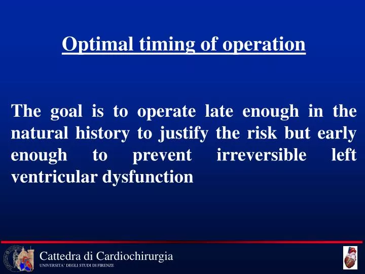
Optimal timing of operation The goal is to operate late enough in the natural history to justify the risk but early enough to prevent irreversible left ventricular dysfunction
Guidelines for the management of Patients with valvular heart disease ACC / AHA presented march 1999 American College of Cardiology 48 Annual Scientific Session
Guidelines for classifying Indications • Class I: Conditions for which there is evidence and/or general agreement that a given procedure or treatment is useful and effective • Class II: Conditions for which there is conflicting evidence and/or a divergence of opinion about the usefulness of a procedure or treatment • II a: Weight of evidence/opinion is in favour of usefulness/efficacy • II b: Usefulness/efficacy is less well established by evidence/opinion • Class III: Conditions for which there is evidence and/or general agreement that the procedure/treatment is not useful and in some cases may be harmful
AORTIC STENOSIS • Mild: aortic valve area > 1.5 cm2 • Moderate: aortic valve area 1.0 - 1.5 cm2 • Severe: aortic valve area < 1.0 cm2 • ( Critical: aortic valve area = 0.75 cm2 )
AS, Rate of progression - Hemodynamic • Cardiac Catheterisation (3-9 year f/u ) • Progression • Valve area decreases = 0.1 - 0.3 cm2 /year • Pressure gradient increases = 10-15 mm Hg/year • Little or no progression in 50% of reported patients • Echocardiography (1-3 year f/u) • Progression • Valve area decreases = 0.1 cm2/year • Pressure gradient increases = 15-19 mm Hg/year • Little or no progression in 50 % of reported patients • Faggiano, et al. Am Heart J 1996
AS, Rate of progression-Symptoms/Need for surgery • Prospective follow up of asymptomatic patients with severe aortic stenosis (Doppler velocity > 4 m/s) • Symptoms developed in 30% within 2 years • Pellikka, et al. JACC 15: 1012, 1990 • Surgery was performed in 70% within 2 years • Otto, et al. Circ 95:2262, 1997
Recommendations for Echo in AS IndicationClass 1 Diagnosis and assessment of severity of AS I 2 Assessment of LV size, function, and or hemodynamics I 3 Reevaluation of patients with known AS with changing I symptoms or signs 4 Assessment of changes in hemodynamic severity and ventricular I compensation in patients with known AS during pregnancy 5 Reevaluation of asymptomatic patients with severe AS I 6 Reevaluation of asymptomatic patients with mild to moderate AS IIa and evidence of LV-dysfunction or hypertrophy 7 Routine reevaluation of asymptomatic adult patients with mild AS III having stable physical signs and normal LV size and function
AS, Exercise Testing • Patient population (n=123) • Asymptomatic adults with AS • Max (Doppler) velocity: average 3.6 m/s • Results (274 tests in 104 patients) • > 80% of max predicted Heart rate in 87% of patients • no morbidity or mortality • BP fell in 25 (9%), eligible for AVR • ST depression in 4 (2%) • Otto, et al. Circ 1997
Recommendations for Catheterizaion in AS IndicationClass 1 CAG before AVR in patients at risk for CAD (see section VIII.B of these I guidelines). 2 Assessment of severity of AS in symptomatic patients when AVR is planned I or when noninvasive tests are inconclusive or there is a discrepancy with clinical findings regarding severity of AS or need for surgery 3 Assessment of severity of AS before AVR when noninvasive tests are IIb adequate and concordant with clinical findings and CAG is not needed 4 Assessment of LV function and severity of AS in asymptomatic patients III when noninvasive tests are adequate
Low-gradient AS • Problem: Low cardiac output and low pressure gradient. Calculated valve area indicates severe stenosis • Determine pressure gradient, valve area/resistance during: • 1 Resting - baseline state • 2 Stress - dobutamine (or exercise) • If dobutamine produces an increment in stroke volume and an increase in valve area, the baseline calculation probably overestimates the severity of the stenosis
Recommendations for AVR in AS 1 IndicationClass 1 Sympomatic patients with severe AS I 2 Patients with severe AS undergoing CABG I 3 Patients with severe AS undergoing surgery of the I aorta or other heart valves 4 Patients with moderate AS (>30) undergoing CABG IIa surgery on the Aorta or other heart valves (see III.F and Viii.D)
Recommendations for AVR in AS 2 IndicationClass 5 Asymptomatic patients with severe AS and . LV systolic dysfunction IIa . Abnormal response to exercise (eg Hypotension) IIa . Ventricular tachycardia IIb . Marked or excessive LVH (>= 15mm) IIb . Valve are < 0.6 cm2 IIb 6 Prevention of SCD in asymptomatic patients with III findings under 5
Recommendations for Balloon Valvulotomy in AS IndicationClass 1 A bridge to surgery in hemodynamically unstable patients IIa who are at high risk for AVR 2 Palliation in patients with serious comorbid conditions IIb 3 Patients who require urgent noncardiac surgery IIb 4 An alternative to AVR III Recommendations for PTVP Ao in adolescents and young adults with AS are provided in VI.A
Mitral Stenosis Etiology Rheumatic fever Leaflet thickening Commissural fusion Chordal fusion
Mitral Stenosis Pathophysiology Narrow Orifice Transmitral Pressure Gradient Halmark of MS Elevated LAP
Mitral Stenosis What is new ? - 2D and doppler echo - Percutaneous Mitral Balloon valvotomy (PMBV) Recommendations for patient care -Asymptomatic -Symptomatic
Mitral Stenosis 2 D echo is the Gold Standard for MS
Mitral Stenosis Doppler echo is the Gold Standard for the quantification of mitral stenosis
Mitral Stenosis Doppler echo is more accurate than conventional catheterization
Mitral Stenosis Percutaneous Mitral Balloon Valvotomy PMBV
PMBV, immediate results Doubling of MVA 50-60 % reduction gradient Success rate 80-95%
Mitral Stenosis Results PMBV Results are even better than for Valve replacement Farhat et al: Circ;97:245-25
Mitral Stenosis • PMBV: Dependent upon mitral morphology • Non calcified, pliable • No commissural fusion • Success > 90% • Complications < 3%
Mitral Stenosis Asymptomatic Mild stenosis MVA > 1.5 cm2 Mod-severe stenosis MVA < 1.5 cm2 Yearly exam ? Suitable for PMBV ?
Mitral Stenosis Asymptomatic ? Suitable for PMBV ? Yes No PAP > 50 PAP < 50 Yearly exam PMBV
Mitral Stenosis Exercise induced pulmonary HTN PMBV Calculated PAP
Mitral Stenosis Symptoms Mild stenosis MVA > 1.5 cm2 Mod-severe stenosis MVA < 1.5 cm2 PAP > 60 Grad > 15 Exercise ? Suitable For PMBV? Pap < 60 Grad<15 yes PMBV Look elsewhere
Mitral Stenosis Symptoms ? Suitable for PMBV ? No Yes Follow Class II Surgery Class III, IV PMBV
Mitral Stenosis • Other issues • Rheumatic fever prophylaxis • Anticoagulation • Treatment for atrial fibrillation • Recommendations for exercise • Pregnancy • Cost-effective follow-up
Aortic Regurgitation • Percent Survival 3 yr after operation for AR: • Pre-op LVEF >= 0.50 : 90 %; Pre-op LVEF < 0.50 : 60 % • Forman et al, Am J Cardiol, 1980 Cheitlin et al Dilemmas in clinical cardiology 1990
Chronic Aortic Regurgitation 1 Preoperative prediction of survival after AVR: Predictor #LVEFLVFSLVSD Forman 1980 90 x Henry 1980 50 x x Gunha 1980 86 x x Greves 1981 45 x Kumpuris 1982 43 x Bonow 1985 80 x x x Daniel 1985 84 x x Cormier 1986 73 x x Shelban 1986 84 x x
Chronic Aortic Regurgitation 2 Preoperative prediction of survival after AVR: Predictor #LVEFLVFSLVSD Taniguchi 1987 62 x x* Klodas 1996 219 x Turina 1998 192 x x* --------------------------------------------------------------------------------------- Total 1108 *LVSV
LV dysfunktion in valvular AR Reversible alteration in LV loading (afterload mismatch) versus Irreversible LV myocardial dysfunction
Chronic AR with LV dysfunktion Factors influencing survival and functional results after AVR: 1 Severity of preoperative symptoms 2 Severity of LV dysfunction 3 Duration of LV dysfunction
Chronic AR with LV dysfunktion Asymptomatic patients with aortic regurgitation and LV dysfunction should undergo operation beforethe onset of symptoms and limitation of exercise capacity
Timing of operation for asymptomatic AR Management considerations: 1 Survival and functional results after aortic valve replacement 2 Natural history of asymptomatic patients
Asymptomatic AR with normal LVF Natural history Rate of progression to symptoms and/or LV dysfunction nRate Bonow, Circ 1984, 1991 104 3.8%/yr Scognamiglio, Clin Cardiol, 1986 30 2.1%/yr Siemenczuk, Ann Int Med 1989 50 4.0%/yr Scognamiglio, N Engl J Med 1994 74 5.7%/yr (+digoxin) Tornos, Am Heart J 1995 101 3.0%/yr Ishii, Am J Cardiol 1996 27 3.6%/yr (incomplete data) Borer, Circ 1998 104 6.2%/yr --------------------------------------------------------------------------- Total 490 4.3%/yr
Asymptomatic AR with normal LVF Natural history Likelihood of developing asymptomatic LV dysfunction nMean F/URate Bonow, Circ 1984, 1991 4/105 8.0 yr 0.5%/yr Scognamiglio, Clin Cardiol, 1986 3/30 4.7 yr 2.1%/yr Siemenczuk, Ann Int Med 1989 1/50 3.7 yr 0.5%/yr Scognamiglio, N Engl J Med 1994 15/74 6.0 yr 3.4%/yr Tornos, Am Heart J 1995 6/101 4.6 yr 1.3%/yr Borer, Circ 1998 7/104 7.3 yr 0.9%/yr -------------------------------------------------------------------------------------- Total 36/463 5.9 yr 1.3%/yr
Asymptomatic AR with normal LVF Event Rate Death < 0.2 % / yr Asymptomatic LV Dysfunction 1.3 % / yr Symptoms and/or LV dysfunction 4.3 % / yr
Asymptomatic AR with normal LVF Factors predictive of symptoms and/or LV dysfunction . LV end systolic dimension/volume . LV end diastolic dimension/volume . LV ejection fraction with exercise Bonow, Circ 1984, 1991 Siemenczuk, Ann Int Med 1989 Tornos, Am Heart J 1995
Asymptomatic AR with normal LVF Likelihood of death, development of symptoms and/or LV dysfunction (Risk Stratification) . LV end systolic dimension/volume > 50 19%/yr 40-49 6%/yr < 50 0%/yr . LV end diastolic dimension/volume >= 70 10%/yr < 70 2%/yr . LVEF response to exercise decrease >5% 12%/yr decrease 0-5% 4%/yr increase > 0% 1%/yr Bonow, Circ 1984, 1991
Asymptomatic AR with normal LVF Predictive variables in multivariate analysis: Initial evaluation: . Age . LV end-systolic dimension Serial evaluation: . Increase in LVSD . Decrease in resting LVEF Bonow et al, Circ 1984,1991
Asymptomatic AR with normal LVF Risk of sudden Cardiac Death: . LV end-diastolic volume > 200 ml/m2 Turina et al, Circ 1984 . LV end-diastolic dimension >= 80 mm . LV end-systolic dimension > 55 mm Bonow et al, Circ 1991
Chronic AR with marked LV dilatation Outcome after AVR: Low risk group: . Asymptomatic with normal EF High risk groups: . Symptoms . LV Dysfunction Klodas et al, JACC 1996, 31 patients with LVDD > =80 mm
Chronic AR Indications for operation: . Symptoms . LV systolic dysfunction (subnormal EF at rest) . Marked LV dilatation (LVSD >= 55 mm; LVDD >= 75mm)
Asymptomatic AR with normal LVF Follow-up strategy . Monitoring for onset of symptoms and changes in effort tolerance . Serial echocardiograms frequency based on LV size and function . Ancillary tests .Exercise treadmill testing if symptoms unclear .Radionuclide angiography if echo data equivocal
Mitral Regurgitation Chronic compensated MI: EDV 240, ESV 50, Filling pressure 15 mm Hg Chronic decompensated MI: EDV 260, ESV 110, Filling pressure 25 mm Hg
Mitral Valve Surgery EF after repair: the same or better EF after replacement: . Chords preserved: the same . Chords severed: worse, sometimes even becomes half of the original value