Download
1 / 1
10 likes | 150 Vues
PROCESSING A POLYELECTROLYTE COMPLEX HYDROGEL AT THE MICRO-SCALE. DANIELA F. COUTINHO* Supervisors: Nuno M. Neves, Manuela E. Gomes * d.coutinho@dep.uminho.pt. INTRODUCTION

E N D
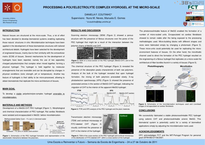
PROCESSING A POLYELECTROLYTE COMPLEX HYDROGEL AT THE MICRO-SCALE DANIELA F. COUTINHO* Supervisors: Nuno M. Neves, Manuela E. Gomes * d.coutinho@dep.uminho.pt INTRODUCTION Natural tissues are structured at the micro-scale. Thus, a lot of effort has been devoted to develop biomaterial systems enabling replicating those natural structures in vitro. Microfabrication techniques have been applied in the development of these biomaterials structures with tailored architectural details. Hydrogels have been selected for the development of engineered tissues, mainly due to their similarity with the extracellular matrix (ECM) of tissues. Several mechanisms for the development of hydrogels have been reported, namely the use of two oppositely charged polyelectrolytes that complex when mixed together, forming a physical hydrogel. This hydrogel is held together by molecular entanglements that are reversible and can be disrupted by changes in physical conditions (ionic strength, pH or temperature). Another key feature of hydrogels is their ability to be micro-processed, allowing to obtain biomaterials that replicate the micro-architecture of tissues. MAIN GOAL To develop a stable polyelectrolyte-complex hydrogel amenable to microfabrication. MATERIALS AND METHODS RESULTS AND DISCUSSION The photocrosslinkable feature of MeGG enabled the formation of a number of micro-scaled units. Encapsulated rat cardiac fibroblasts showed to remain viable after the being exposed to the processing methodologies used. Micro-building blocks with different shapes and sizes were fabricated simply by changing a photomask (Figure 5). These micro-units could potentially be used for replicating the micro-environment features of tissues. On the other hand, the microfluidic channel used to direct the formation of the PEC hydrogel resulted on the engineering of a fibrous hydrogel that replicates at a micro-scale the architecture of fiber bundles found in a variety of tissues (Figure 5). Scanning electron microscopy (SEM) (Figure 2) showed a porous structure with the presence of fibrous structures over the pores of the PEC hydrogel that might be a result of the interaction between the polymer chains of MeGG and CHT. Figure 2. SEM of a cross-section of the PEC hydrogel (MeGG-CHT) and of the plain materials. The chemical structure of the PEC hydrogel (Figure 3) revealed the presence of the absorption peaks characteristic of both raw polymers. Analysis of the bulk of the hydrogel revealed that upon hydrogel formation, the mixing of both polymers proceeded slowly. X-ray photoelectron spectroscopy (XPS) (Figure 3) showed the presence of protonated amines in the section of MeGG-CHT hydrogel, indicating the migration of CHT to the interior of the apparent MeGG hydrogel. Photolithography Microfluidics FTIR XPS CHT MeGG-CHT MeGG-CHT section Figure 5. Schematics of the microfabrication techniques used and Live/dead images of encapsulated rat cardiac fibroblasts Development of a MeGG-CHT PEC hydrogel (Figure 1). Morphological and chemical characterization of PEC hydrogel. Rat cardiac fibroblasts were isolated and encapasulated in MeGG before microfabrication. Figure 3. FTIR and XPS analysis of the PEC hydrogel and the plain materials. CONCLUSIONS We successfully fabricated a stable photocrosslinkable PEC hydrogel using cationic CHT and photocrosslinkable anionic MeGG. This biomaterial system is potentially useful for a variety of biomedical applications simply by adjusting the microfabrication tools used. TEM Confocal Transmission electron microscopy (TEM) and confocal microscopy of FITC-labeled CHT (Figure 4) visually confirmed the presence of CHT in the interior of the hydrogel. Methacrylated Gellan Gum - 1% (w/v) in dionized water MeGG-CHT PEC hydrogel 1st - Hydrogelformedthroughelectrostaticinteractions 2nd - Hydrogelstabilizedby UV ACKNOWLEDGEMENTS DFC acknowledges FCT and the MIT-Portugal Program for personal grant SFRH/BD/37156/2007. Chitosan - 1% (w/v) in acetic acid solution Figure 4. TEM of the cross-section of the PEC hydrogel and confocal microscopy of the distribution of FITC-labeled CHT within a cross-section of the hydrogel. Figure 1. Schematics of the PEC hydrogel formation and representation of the electrostatic interactions between the polymers.