Download
1 / 61
720 likes | 1.23k Vues
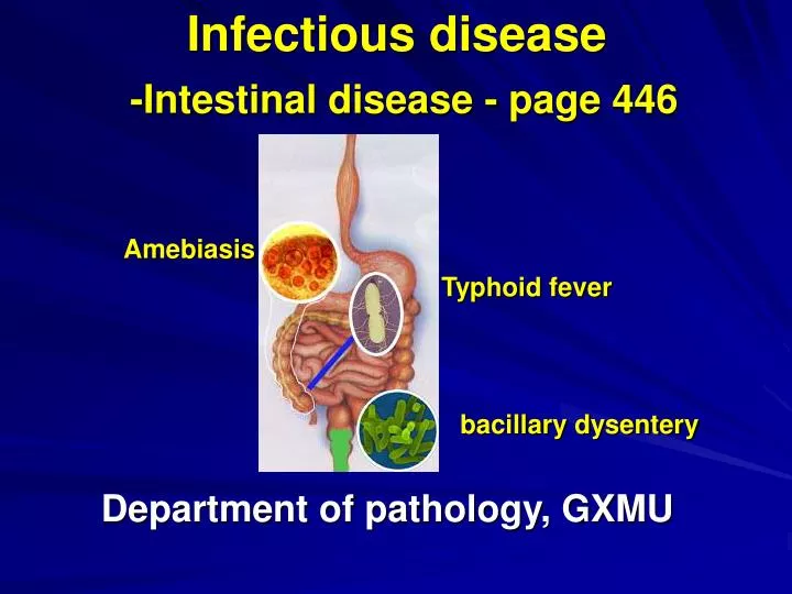
Infectious disease -Intestinal disease - page 446. Amebiasis. Typhoid fever. Department of pathology, GXMU. bacillary dysentery. Introduction. Infectious source: patients & healthy carriers Transmit Pattern: Fecal-oral route Pathogen: bacilli or parasites. Key points. Inflammation type

E N D
Infectious disease-Intestinal disease - page 446 Amebiasis Typhoid fever Department of pathology, GXMU bacillary dysentery
Introduction Infectious source: patients & healthy carriers Transmit Pattern: Fecal-oral route Pathogen: bacilli or parasites
Key points Inflammation type Most common location Intestinal ulcer Diarrhea
Typhoid fever 公元3世纪之初期,张仲景博览群书,广采众方,凝聚毕生心血,写就《伤寒杂病论》。伤寒,又谓,“伤于寒邪”, 中医所说的伤寒,广义上指的是外感热病的总称,狭义指的是外感风寒之邪,感而即发的疾病。
Typhoid fever- Introduction • Thomas Willis can be regarded as the pioneer in typhoid fever. Until his classic description in 1659 and its translation into English in 1684, little had been done to separate this disease from the other disease with fever. • A systemic infection disease presenting as continued fever with relative badycardia ,and abdominal symptoms and psychosis confusion. • Is characterized by involvement of mononuclear phagocytic system (MPS), with typhoid nodule formation, especially in the Peyer’s patches and solitary lymph follicles of lower ileum.
Epidemiology • Is still a very important problem in many developing countries • 17 Million cases occur per year worldwide, 7 million distribute in Asia, 4 million in Africa, 0.5 million in Latin America • Can be prevented by vanccine High population densities and poor sanitation Distribution
Pathogen • Salmonella typhi (typhoid bacilli) • typhoid bacilli are rod-shape, 2-3um long and 0.4-0.6 um in diameter • Three antigenic structures: O antigens; H antigens; Vi antigens • Endotoxin Widal reaction flagellum
Transmission • Patients • Healthy carriers (“Typhoid Mary” ) • Fecal-oral pattern • Flies
Tyhpoid Mary in the cartoons Meet the ladies who drive heroes crazy. These are the women you kill for, the women you die for.
Tyhpoid Mary in the movie "Elektra"Movie Photo (Center) Natassia Malthe (Typhoid Mary) and Will Yun Lee (Kirigi) in 20th Century Fox's "Elektra." - A generic term for a carrier of a dangerous disease who is a danger to public because they refuse to take apporpriate precaution
Ingestion Pathogenesis Invade the mucosa Taken up by macrophages and transported to regional lymph node Incubation period – the first week multiplies in lymphoid tissue Bacteremia phase Systemic illness – at the end of the first week Toxemia phase Reinfects lymphoid tissue –endotoxin and delayed hypersensitivity reaction Intestinal illness – the second week
Pathological Changes Typhoid Cells Typhoid nodule (granuloma) formation ? • It is a localized accumulation of large mononuclear cells • such as rheumatism, tuberculosis
Typhoid cell Erythrophagocytosis Erythrophagocytosis Typhoid cells and typhoid nodules (granuloma) is hallmark histologic finding in typhoid fever
Typhoid nodule formation in the liver Typhoid cells and typhoid nodules (granuloma) is hallmark histologic finding in typhoid fever
Intestinal lesion • Lower ileum and cecum • 4 stages (last 4 weeks) Hyperplasis of Peyer’s pathes Necrosis Ulceration Healing
Peyer’s patches: a collection of lymphoid follicles Locate in mucosa and extend into sumucosa Terminal ileum contains most peyer’s patches
1st Stage (First week): Hyperplasia of peyer’s pathes The phagocytes in Peyer’s patches of the ileum and the solitary lymph follicles are proliferation and Hyperplasia Macroscopilly Projected on the mucosal surface Microscopilly Typhoid granuloma with a large number of typhoid cells can be seen obviously Clinical Blood culture † † † † Stool culture -
2nd Stage(The second week) : Necrosis • Yellow or greenish-brown • From center to peripheral Clinical Blood culture † † † Stool culture †
3rd Stage(The third week) : Ulceration Rounded or oval , deep ulcer,which long axis is in the direction of the long axis of the bowel (Longitudinal ulcer-typical finding of typhoid by macroscopically). Compare: Transverse Ulcer? Clinical Stool culture ††† Widal reaction †††
4th Stage (The fourth week): Healing Clinical Widal reaction ††††
Extraintestinal Lesions Phagocytes proliferate in Reticuloendothelial system(网状内皮系统, mononuclear phagocytic system ) • Spleen (Sinus histiocytes) -Splenomegaly • Liver (Kupffer) - hepatomegaly • Lymph nodes • Bone marrow The reaction tends to be similar everywhere, with proliferation of large mononuclear cells and foci of necrosis
Clinical features • Bacteremia: blood culture / stool and urine culture • Toxemia : Disorientation, delirium(暂时精神乱),Restleness,Headache , Rose spots,Continued fever(稽留热) ,diarrhea ,Relative Bradycardia • Splenomegaly and/or hepatomegaly • Leukopenia(白细胞少症状) Complications Hemorrhage Perforation
Days 1st Stage 2nd Stage 3rd Stage 3th Stage Stage Rose spots Splenomagly Psychosis confusion Leukopenia Blood culture Stool culture Widal reaction Continued fever(稽留热) and Relative Bradycardia: the classical type of pyrexia with its step-ladder rise during the first week, its maintenance during the second and third weeks, and its fall in the fourth week
rose spots:2-4mm in diameter appear on the trunk of patients;
Summary 1 Pathogen: Salmonella typhi 2 Inflammation: Granuloma 3 Pathological stages: Longitudinal ulcer 4 Clinical features: Continued fever, diarrhea, relative badycardia , abdominal symptoms and psychosis confusion
Bacillary dysentery Introduction is an acute infectious inflammatory disease of the colon caused by Shigella bacteria; characterized by bloody mucoid diarrhea, tenesmus(里急后重) and abdominal pains. It commonly occurs in summer and fall.
Etiology and pathogenesis Four species of Shigella: S.Flexneri 福氏 S.Sonnei (the most comon cause) 宋内氏 S.Boydii 鲍氏 S.Dysenteriae 志贺氏 -Minimal infective dose is less than 1000 organisms Endotoxin • Patients • Healthy carriers • Fecal-oral route
Pathological changes and clinical types • Location: large intestine, sigmoid, rectum(only involve the superficial layer) • Three types Acute bacillary dysentery Chronic bacillary dysentery Toxic bacillary dysentery
Acute bacillary dysentery congestion Acute catarrhal inflammation edema infiltration Acute pseudomembranous inflammation (Fibrinous inflammation) pseudomembrane Lyse fall off Irregular,map-like,shallow ulcers
A pseudomembrane covered on the mucosal surface , yellowish or yellow-greenish in color
Plaques of yellow fibrin and inflammatory debris are adherent to a reddened colon mucosa..
Bloody mucoid Diarrhea?Tenesmus? Pseudomembrane Mucosa Submucosa The Pseudomembrane consist of a large deal of fibrin, necrotic epithelium, neutruphils, RBC and bacteria. but the submucosa isn’t greatly involved.
Shallow, irregular, ragged Map-like ulcers Superficial scar formation Stenosis ,hemorrhage and perforation are uncommon Compare: Transverse Ulcer and longitudinal ulcer
Passage of 10-40 stools per day is usual,stools compose of blood,mucus and neutrophilics Clinical features • Bloody mucoid diarrhea are more commonly • Abdominal discomfort and tenesmus • Fever,headache,tireness and anorexia(食欲减退) • May last 1 to 2 weeks
Chronic bacillary dysentery • Transformed from acute bacillary dysentery. • The clinical course exceed 2 monthes • S.flexneri infection are more common • Pseudomembrane,Ulcerations(new lesions) and granulation tissue organization(old lesions) progress repeatedly • Polypi formation & stenosis of the bowel occur • Bacillary culture from stool is persistent positive
Toxic bacillary dysentery • 2~7y children • S.flexneri & S.sonnei infection • Intestinal lesions are mild while general toxic symptoms are severe • Toxic shock & breath failure occur rapidly
Complications • Bacteremia and septicemia (Malnourished children) • Hemolytic uremic syndrome (溶血性尿毒症综合征) • Central nervous system lesions • Myocarditis
Summary 1 Pathogen: Shigella bacteria 2 Inflammation: Fibrinous inflammation 3 Pathological stages: Map-like ulcer 4 Clinical features: Bloody mucoid diarrhea and tenesmus
Amebiasis Introduction • Refers to the infection caused by Entamoeba histolytica • Transmit: Fecal-oral route • May penetrate the mucosa and possibly invade locally(Intestinal amebiasis) or by hematogenous spread to other organs such as liver, lung, brain and cause the organs of amebic abscesse • Human beings are the only known host of the ameba
Epidemiology • Worldwide distribution • 50 million new cases annually; • 50 to 100 thousand deaths among them • Higher morbidity in rural area • Risk groups Travelers, recent immigrants are most at risk
Intestinal amebiasis • An infection disease caused by E. histolytica that inhabits the intestinal tract. • Fecal-Oral transmission • Diarrhea - -- Amebic dysentery • Low grade fever
Ingestion of cysts Etiology and pathogen intestinal alkaline medium Encysted organism becomes small trophozoites Small trophozoites develop adult trophozoites Move to cecum become a commensal or a highly invasive pathogen Lyse host tissue Become cysts
Pathogenesis of Amebiasis • Adult Trophozoites ... • Attach to mucosal epithelial cells (MEC) • Lyse MEC (contact-dependent cytolytic mechanism) • Ulcerate and invade mucosa • Cause dysentery • spread to other organs via blood to cause a Amebic abscesses in extraintestinal sites Contact lyse by enzyme or Enzymatic necrosis
Pathology Location: Most frequently in cecum, less frequency in the ascending colon, sigmoid, rectum, and appendix. Pinpoint-sized ulcers Fastener-shaped ulcers Flask-shaped ulcers Large, undermined edges ulcers Ulcer formation:
mucosa mouth neck bottom submucosa Flask-shaped ulcer
Amoebae are found in the base and at the margins of ulcers, chiefly in the submucosa