Download
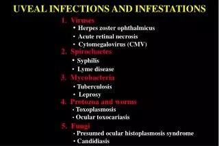
1 / 48
510 likes | 987 Vues
Uveal Diseases. Umut Aslı Dinç., MD., FEBO Associate Professor in Ophthalmology Yeditepe University Eye Hospital. Uveal Tract. Pigmented, vascular structure that lies between the sclera&retina Consists of: İris Ciliary body Choroid. Uveal Tract.

E N D
Uveal Diseases Umut Aslı Dinç., MD., FEBO Associate Professor in Ophthalmology Yeditepe University Eye Hospital
Uveal Tract Pigmented, vascular structure that lies between the sclera&retina Consists of: • İris • Ciliary body • Choroid
Uveal Tract • Supplies most of the ocular vasculature through the anterior and posterior ciliary branches of ophthalmic artery. • Produces aqeous humor • Controls accomodation at near • Supplies aqeous outflow by trabecular meshwork
Uveitis • Inflammation of the uveal tract and adjacent structures. • Mostly the cause is unknown.
Uveitis Classification • Anterior uveitis (iris and ciliary body) Iritis, anterior cyclitis, iridocyclitis • Intermediate uveitis (pars plana of ciliary body, anterior vitreus and peripheral retina) Posterior cyclitis, pars planitis • Posterior uveitis (choroid) Choroiditis, chorioretinits • Panuveitis
Uveitis Classification • Acute uveitis • Chronic uveitis
Acute Uveitis-Clinical Features • Pain • Redness • Photophia • Epiphora • Blurred vision • Floaters
Chronic Uveitis-Clinical Features • Fewer or none of the acute symptoms • Periods of exacerbations and remissions
Causes of Uveitis • Idiopathic • Infectious (bacterial, viral, fungal, parasitic) • Traumatic • Post-surgery • Tuberculosis • Sarcoidosis • Behçet’s disease • Spondiloartropathies • Inflammatory bowel diseases • Collagen vascular diseases • Medication
History taking in uveitis • Present illness Onset, course, symptoms, laterality • Past ocular history Previous episodes, treatment, ocular trauma or surgery • Medical history Systemic disease (sarcoidosis, tuberculosis, syphilis, Juvenile rheumatoid artritis, AIDS, etc), maternal infection • Sexual history, intravenous drug abuse • Demograhic data Age, sex, race
History taking in uveitis Review of symptoms • General-fever, wegit loss, malaise, night sweats • Rheumatologic-arthralgias, lower back pain, joint sitffness • Dermatologic-rashes, sores, alopecia, vitiligo, poliosis, insect bites • Neurologic-tinnitus, headache, meningism, paresthesias, weaksness/paralysis • GIS- diarrhea, bloody stools, aphtous ulcers • GUS-dysuria, discharge, genital ulcers, balanitis
Uveitis-Clinical Features • Inflammatory cells in the anterior chamber • Keratic precipitates on corneal endothelium • Anterior synechiae (adhesions of iris to cornea) • Posterior synechiae (adhesions of iris to lens) • Inflammatory cells in the vitreous cavity (vitritis) • Sheating of retinal vessels • Optic disc or macular edema • Choroidal or chorioretinal infiltrates
Anterior Uveitis • Anterior segment cells • Anterior segment flare
Anterior Uveitis Scleral injection Keratic precipitates on corneal endothelium
Anterior Uveitis • Posterior synechiae
Anterior Uveitis • Idiopathic • Infectious (Herpetic uveitis, Bacteriel uveitis) • Traumatic • Fuch’s heterochromic iridocyclitis • Immune-mediated Behçet’s disease Seronegative spondiloartropathies (Ankylosing Spondylitis, Reactive (Reiter) Arthritis, Psoriatic Arthritis ) Inflammatory bowel diseases (Crohn disease, ulcerative colitis) Juvenile Rheumatoid Arthritis • Sarcoidosis • Tuberculosis • Toxic (Rifabutin, sulfonamides, cidofovir)
Idiopathic anterior uveitis • Most common form of ocular inflammation • No systemic or ocular cause • Relief with topical steroids and cycloplegic drops
Fuch’s heterochromic iridocyclitis • Unilateral • Heterochromia (lighter iris color is typical) • Vision loss secondary glaucoma and cataract
Fuch’s heterochromic iridocyclitis Heterochromia due to iris atrophy
Herpetic uveitis • Keratitis • Iris atrophy
Behçet’s disease Key features • Uveitis (anterior, posterior) • Recurrent oral ulcers • Recurrent genital ulcers • Skin lesions Associated features • Erythema nodosum • Arthritis • İntestinal ulcers • Vascular lesions-thrombophlebitis, arteriel occlusions, aneurysms • CNS involvement
Behçet’s disease • HLA-B51 • Pathergy test +
Seronegative spondiloartropathies • HLA B27 + • Sacroiliac joint radiography
Juvenile Rheumathoid Arthritis • Most frequently in RF (-), ANA (+), oligoarticular type • White eye • Band keratopathy • Posterior synechiae • Cataract
Sarcoidosis • Noncaseating granuloma • Anterior and posterior uveitis • Bilateral hilar lymphadenopathy • Pulmonary parenchymal disease • Chest radiography • Serum ACE enzyme
Sarcoidosis • Granulomatous reaction with iris nodules and mutton fat keratic precipitates
Tuberculosis • Caseating granuloma • Anterior and posterior uveitis • Chronic inflammation • PPD test • Chest Radiography
Tuberculosis • Granulomatous reaction with iris nodules and mutton fat keratic precipitates
Intermediate Uveitis • Blurred vision and floaters • Typically bilateral • Vision loss secondary to cystoid macular edema • Pars planitis... idiopathic type
Intermediate Uveitis • Vitreous snowballs • Snowbanking (exudation at pars plana)
Intermediate Uveitis Perivascular sheating Vitreous snowball
Posterior Uveitis • Vitritis • Choroiditis • Retinitis • Papillitis • Retinal detachment
Posterior Uveitis • Viral (CMV, VZV, HIV in immunodeficiency) • Bacterial • Fungal (candida albicans, histoplasma capsulatum, coccidioides immitis) • Toxoplasmosis • Parasitic (toxocariasis, cysticercosis, onchocerciasis) • Syphilis • Behçet’s disease • Sarcoidosis • Tuberculosis • Syphilis
Viral Posterior Uveitis CMV chorioretinitis
Viral Posterior Uveitis Acute retinal necrosis secondary to HSV infection
Viral Posterior Uveitis HIV chorioretinitis
Fungal Posterior Uveitis Ocular candidiasis
Toxoplasmosis • Toxoplasma gondii • Transmission by infected raw meat or congenitally via plasenta • Recurrent chorioretinitis and severe vitritis • Toxoplasma IgM, IgG and PCR
Congenital Toxoplasmosis Nonactive chorioretinal scars
Toxoplasmosis “Headlight in fog”
Toxocariasis • Typical toxocara granuloma • Traction of macula, optic disc • Tx: steroids • Severe inflammation when microorganism dies.
Tuberculosis Choroidal tubercule
Syphilis Usually in acquired syphilis VDRL, RPR, FTA-ABs+
Anti-inflammatory Therapy • Corticosteroids Topical drops/ periocular injection/ systemic) • Cytotoxic drugs Antimetabolites (azathioprine, methotrexate) Alkylating agents (cyclophoshamide, chlorambucil) • Immunomodulator agents (cyclosporin, tacrolimus) • Anti-TNF agents (etanercept, infliximab, adalimumab)
Complications of Uveitis • Band keratopathy • Posterior synechiae • Cataract • Glaucoma • Cystoid macular edema • Optic atrophy