Download

1 / 40
780 likes | 1.22k Vues
NAEGLERIA FOWLERI. BRAIN EATING AMOEBA AMOEBIC MENINGOENCEPHALITIS. DR PRIYADARSHEE PATEL Medical Graduate INDIA NEUROLOGY. PARASITE. Amoeba - Protozoa, Genus Naegleria, P hyl lum Percolozoa Thermophillic F ree-livin g amoeba

E N D
NAEGLERIA FOWLERI BRAIN EATING AMOEBA AMOEBIC MENINGOENCEPHALITIS DR PRIYADARSHEE PATEL Medical Graduate INDIA NEUROLOGY
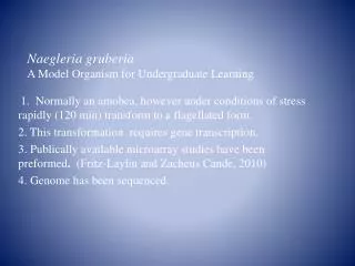
PARASITE • Amoeba- Protozoa, Genus Naegleria, Phyllum Percolozoa • Thermophillic • Free-living amoeba • Causes Fulminant (sudden and severe) brain infection called Naegleriasis (Primary amoebic meningoencephalitis)
PARASITE • Found in warm freshwater bodies • Ponds, lakes, rivers, hot springs • Man made waterbodies • Unchlorinated swimming pools • Soil near discharges of industrial plants
PARASITE • Sensitive to harsh environment • Airidity • Extreme Ph • Sea water • Feeds on • Bacteria • Microorganisms • Brain tissues
The life cycle of the ameba has 3 stages: Trophozoite Flagellar Cyst LIFE CYCLE
THE TROPHOZOITE Vegetative or feeding stage of the ameba. Measures 10-20 µm in diameter Agranular cytoplasm and a distinct ectoplasm. Actively motile with the help of a broadly rounded, granule-free projectionoriginating from the surface. Helps to ingest bacteria, yeast cells, and cellular debris and may serve as an organelle of attachment. In tissue, trophozoites ingest red and white blood cells and cause tissue destruction. The trophozoite stage is the only one in which the ameba multiplies via binary fission.
THE FLAGELLATE Temporary form of the ameba in which it neither feeds nor divides in culture. The ameba progresses to the amebo-flagellate stage when the trophozoites form is exposed to a change in ionic concentration, such as in distilled water. During the amebo-flagellate stage, the parasite is pear-shaped with a flagellar apparatus at the broader end.The flagellar apparatus consists of 2 terminal flagella, 2 basal bodies, microtubules, and a single striated rootlet, or rhizoplast. May exhibit a rapid forward movement or a slowly spinning circular movement. It reverts to the trophozoite stage within 24 hours.
THE CYST Resistant form of the parasite, offering protection from desiccation and food shortage. Round. Measures 7-10 µm in diameter Surrounded by a smooth double-layered 1-µm wall. Single nucleus, contractile vacuoles, and food vacuoles. Cysts are usually absent in clinical specimens, as the infection is so rapid and fatal that the patient typically dies before the trophozoites encyst.
EPIDEMIOLOGY • The first PAM infection was reported in 1965 in Australia. • From 1965 to 2015 all over world 560 cases have been reported with only 10 survivers. • The risk of infection has been estimated at 1 case per 2.6 million exposures to N. fowleri. • Majority of cases all over world have been reported in the United States. • Cases have been reported in the Czech Republic, Australia, Mexico, New Zealand, Nigeria, Great Britain,India, Pakistan, Malasiya and Taiwan. • Cases of PAM are on rise recently and it is emerging as an emergency medical condition.
In the United States there have been 138 PAM infections from 1965 to 2015 with only four survivors. These infections are primarily reportedmostlyin southern states Majority in Texas and Florida Recentely PAM infections are also reported from northern states as Minnesota, Kansas, Virginia A possibility of a change in trend in the epidemiology of PAM in the United States is suggested. EPIDEMIOLOGY
HOW YOU GET INFECTED? • Swimming • Diving • Playin in mud • Water activities • Tape water • Neti pots • Feces of animals
PATHOPHYSIOLOGY • N. Fowleri trophozoit enters in humanbody through nose while swimming or diving in warm waters contaminated with the parasite. • Swallowing the contaminated water does not cause PAM. • Invades its host by penetrating the olfactory mucosa. • During the initial stages of infection, the host response is initiated by the secretion of mucus that traps the trophozoites.
PATHOPHYSIOLOGY • Despite this response, some trophozoites are able to reach, adhere to, and penetrate the olfactory epithelium because of 37 kda mucinolytic protein • After invading the olfactory mucosa and bulbs, penetrate the submucosal nervous plexus, invade the cribriform plate, and reach the subarachnoid space. • Glucose and protein in the CSF support the growth and multiplication of the amebae. • The high content of oxygen in the CSF and in the brain also facilitates growth of the amebae.
PATHOPHYSIOLOGY • The trophozoites enter the ventricular system through the foramen of Luschka and Magendie and reach the choroid plexus. These then destroy the ependymal layer of the third, fourth, and lateral ventricles and produce acute ependymitis. • They multiply by a process known as promitosis, during which an intact nuclear membrane is present.
PATHOGENICITY • N. fowleri can cause a lethal infection of the brain called Naegleriasis- Primary Amoebic Meningoencephalitis (PAM) • Human to Human transmission is not reported • Incubation Period- 9 15 days • Once the trophozoites ingest brain tissue, symptoms begin to appear • Death will usually occur in 2 weeks
CLINICAL FEATURES Clinical presentation is similar to Bacterial Meningitis with very rapid progress This is the reason of delayed diagnosis and specific management Leading to Fatal outcome
SYMPTOMS • Sever Frontal Headache • Fever • Nausea, Vomiting • Alteration of smell and taste • Photophobia • Nose bleeds • Confusion • Loss of balance • Euphoria • Hallucinations • Seizures
SIGNS • Fever • Stiff neck • Kerning, Brudzinski positive • Swollen lymph nodes • Cranial Nerve Palsy • Altered mental status • Rapid and shallow breathing • Coma
INVESTIGATIONS • Clue to Survival is totally based on early diagnosis • Rarity of the infection and difficulty in initial detection, about 75% of diagnoses are made after the death of the patient • Only in few laboratories in US, Specific tests for N. Fowleri is carried out
INVESTIGATIONS • Diagnostic Work up depends on identification of motile trophozoites on staining and serological testing to identify antigen and nucleic acid • CSF Sample, Brain Biopsy
INVESTIGATIONS • LP Puncture CSF Analysis • Findings are similar to Acute Bacterial Meningitis with Gram stain Negative • Staining to visualize fast motile trophozoits • Serological studies to identify N Fowleri antigen, nuclic acid • Direct visualization • PCR • Antigen detection • Amoebic culture
INVESTIGATIONS • Imaging features of PAM are nonspecific • Findings of CT and MR imaging may be normal early in the disease • Specific Findings: • Brain edema and basilar meningeal enhancement • Hydrocephalus • Obliteration of the cisterns with enhancing basilar exudates • Infarction of the right basal ganglia, due to obliteration of the perforating vessels by the extensive exudates.
INVESTIGATION • Autopsy findings • Pathologic changes in cases of PAM are typified by the extensive damage to the brain parenchyma, ependyma, andmeninges. • Congestion of the meningeal vessels, edematous cortex with herniation of uncus and cerebellum
INVESTIGATION • Microscopically, there is a purulent leptomeningeal exudate with hemorrhage and necrosis throughout the cerebral hemispheres, brain stem, cerebellum, and upper spinal cord.
INVESTIGATIONS • Environmental Detection: Water samples can be collected, concentrated, and put into culture to grow and select for Naegleria fowleri. Samples can be tested using the serologic or molecular methods described above.
TREATMENT • Early diagnosis, treatment, and aggressive supportive care hold the only chance for survival in patients with Primary amebic meningoencephalitis (PAM) • Amphotericin B • Primary treatment for naegleria infection • Antifungal drug,usually given intravenously or intrathecally to kill the amoebas • Mechanism of action- Ultrastructural examination of amebae treated with amphotericin B revealed membrane distortions, including the nuclear envelope, rough and smooth endoplasmic reticula, and plasma membrane blebbing. • Minimum amebacidal concentrations of amphotericin B were determined to be 0.02–0.078 µg/mL. • Side effects ranging from headache to seizures hypokelemia and cardiac arrythmia
TREATMENT • Miltefosine- investigational drug, used for breast cancer and anti-leishmania drug • Mechanism of action- Interacts with membrane lipids, inhibition of cytochrome c and apoptosis of cell • In conjunction with the FDA, the CDC has an expanded access investigational new drug (IND) protocol in effect to make miltefosine available directly from the CDC for treatment of free-living amebae (FLA) in the United State • Chlorpromazine
TREATMENT • Corifungin a novel macrolide, with higher activity against Naegleria than amphotericin B. • In vitro and in vivo animal studies, the authors have demonstrated the superiority of corifungin over amphotericin B. Based on these results, corifungin has been given an orphan drug status by the US Food and Drug Administration
TREATMENT • Hypothermia to prevent further brain damage • Neurosurgical interventions such as Ventriculotomy as a emergency procedure to relieve increased intracranial presure and prevent brain herniation • Intravenous and Intrathecal Miconazole, Clotrimazole, Itraconazole, Fluconazole, Ketoconazole, and oral Rifampicine with varying degrees of efficacy
PREVENTION • Amoebic Meningoencephalitis is 95% FATAL, but 100% PREVENTABLE • Refrain from water related activities during summer months June to September • Avoid swimming or jumping in warm fresh waterbodies • Hold your nose shut, use nose clips or plugs when jumping or diving into water • Use earplugs, swim goggles, or masks if you tend to get ear or eye infections. • Wash open skin cuts and scrapes with clean water and soap. • Avoid digging in, or stirring up, the sediment while taking part in water-related activities in shallow, warm freshwater areas. • Avoid putting your head under the water in hot springs
A Non-Government organization, founded by Jeremy and Julie Lewis of Arlington, Texas after they lost their son Kyle to the disease in 2010 PREVENTION
CASE 1 In late August, the previously healthy boy was evaluated in a local emergency department for a 2-day history of headache and emesis; he was febrile and lethargic without focal neurologic or meningeal signs. CT scan of the head without contrast was normal. Lumbar puncture was unsuccessful Started on intravenous antibiotics for suspected bacterial meningitis. Within several hours of admission, he had spontaneous nonpurposeful movements, was unable to follow verbal commands, and was transferred to a children's hospital intensive care unit (ICU). En route to the ICU, he had a 30-minute right-sided seizure.
CASE 1 A CT scan of the head on admission to the ICU showed edema of the midbrain MRI demonstrated areas of meningeal enhancement in the brainstem suggestive of meningitis. No organisms were observed on a Gram-stained smear of cerebrospinal fluid (CSF) CSF antigen-detection tests were negative for bacterial pathogens. Fresh preparation of CSF revealed no amebae. CSF red blood cell count was 1,550/mm3 (normal: 0/mm3), white blood cell count was 13,650/mm3 (normal: 0--5/mm3), glucose was <5 mg/dL (normal: 40--70 mg/dL), and protein was 679 mg/dL (normal: 12--60 mg/dL).
CASE 1 Follow-up lumbar puncture later the same day revealed motile amebae in a centrifuged CSF specimen. The patient was started on intravenous amphotericin and oral rifampin and ketoconazole
CASE 1 Approximately 12 hours after admission to the ICU, the patient had apneic episodes and anisocoria and was tracheally intubated. Treatment included hyperventilation, hypertonic sodium chloride infusion, mannitol infusion, and the placement of a ventriculostomy. Despite these efforts, the patient's condition worsened, with progressive neurologic deterioration. On the fourth hospital day, the patient died. A postmortem lumbar puncture demonstrated a few motile amebae
CASE 2 • After 35 years without a Naegleria survivor in the United States, during the summer of 2013, a 12-year-old girl, was diagnosed with PAM approximately 30 hours after becoming ill and was started on the recommended treatment within 36 hours. • She also received the investigational drug miltefosine, and her brain swelling was aggressively managed with treatments that included therapeutic hypothermia. • This patient made a full neurologic recovery and returned to school. Her recovery has been attributed to early diagnosis and treatment and novel therapeutics including miltefosine and hypothermia .
ANY SUSPECTED CASE OF PRIMARY AMOEBIC MENINGOENCEPHALITIS MUST BE REPORTED TOCenter of Disease control and prevention