Download
1 / 32
360 likes | 494 Vues
Post-transcriptional Modification. Md. Habibur Rahaman (HbR) Dept. of Biochemistry and Microbiology North South University. Pre-mRNA Processing. In bacterial cells , transcription and translation take place simultaneously

E N D
Post-transcriptional Modification Md. Habibur Rahaman (HbR) Dept. of Biochemistry and Microbiology North South University
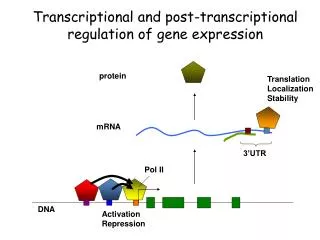
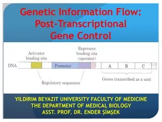
Pre-mRNA Processing In bacterial cells, transcription and translation take place simultaneously while the 3‘ end of an mRNA is undergoing transcription, ribosomes attach to the Shine-Dalgarno sequence near the 5‘ end and begin translation. little opportunity for the bacterial mRNA to be modified before protein synthesis. • In eukaryotic cells: • transcription and translation are separated in both time and space • Transcription takes place in the nucleus, whereas most translation takes place in the cytoplasm; this separation provides an opportunity for eukaryotic RNA to be modified before it is translated.
Eukaryotic mRNA is extensively altered after transcription. Changes are made to the 5‘ end, the 3‘ end, and the protein-coding section of the RNA molecule. The initial transcript of protein-encoding genes of eukaryotic cells is called pre- mRNA, whereas the mature, processed transcript is mRNA. We will reserve the term mRNA for RNA molecules that have been completely processed and are ready to undergo translation.
The Addition of the 5’ Cap Almost all eukaryotic pre-mRNAs are modified at their 5‘ ends by the addition of a structure called a 5‘ cap. This capping consists of the addition of an extra nucleotide at the 5‘ end of the mRNA and methylation by the addition of a methyl group (CH3 ) to the base in the newly added neucleotide and to the 2‘–OH group of the sugar of one or more nucleotides at the 5‘ end. Capping takes place rapidly after the initiation of transcription and the 5‘ cap functions in the initiation of translation.
Early in the elongation process, the 5’ ends of eukaryotic pre-mRNAs are modified by the addition of 7-methylguanosine (7-MG) caps by three enzymatic steps These 7-MG caps are added when the growing RNA chains are only about 30 nucleotides long.
three phosphates are present at the 5’ end of all RNA molecules The 5’ end of pre-mRNA can be represented as 5‘–pppNpNpN, in which the letter N represents a ribonucleotide and p represents a phosphate. Shortly after the initiation of transcription, one of these phosphates is removed and a guanine nucleotide is added
This guanine nucleotide is attached to the pre-mRNA by a unique 5’–5’ triphosphate linkage, which is quite different from the usual 5’–3’ phosphodiester bond that joins all the other nucleotides in RNA.
One or more methyl groups are then added to the 5’ end; the first of these methyl groups is added to position 7 of the base of the terminal guanine nucleotide, making the base 7-methylguanine. Next, a methyl group may be added to the 2’ position of the sugar in the second and third nucleotides. Rarely, additional methyl groups may be attached to the bases of the second and third nucleotides of the pre-mRNA.
Cap-binding proteins recognize the cap and attach to it; a ribosome then binds to these proteins and moves downstream along the mRNA until the start codon is reached and translation begins. The presence of a 5’ cap also increases the stability of mRNA and influences the removal of introns.
The Addition of the 3’-Poly(A) Tail Most mature eukaryotic mRNAs have from 50 to 250 adenine nucleotides at the 3’ end (a poly(A) tail). These nucleotides are not encoded in the DNA but are added after transcriptionin a process termed polyadenylation.
RNA Splicing The other major type of modification that takes place in eukaryotic pre-mRNA is the removal of introns by RNAsplicing. This occurs in the nucleus following transcription but before the RNA moves to the cytoplasm
Fewer, but still many of the genes of multicellular eukaryotes such as the yeasts contain noncoding introns. • Rare genes of a few viruses of prokaryotes and an archebacteria also contain introns. • In the case of these “split gene”, with coding sequences interrupted by noncoding sequences, the primary transcript contains the entire sequence of the gene and noncoding sequences are spliced out during RNA processing. An interrupted gene (also called a split gene) is a gene that contains sections of DNA called exons, which are expressed as RNA and protein, interrupted by sections of DNA called introns, which are not expressed. The DNA sequence in the exon provides instructions for coding proteins.
For genes that encode proteins, the splicing mechanism must be precise; it must join exon sequences with accuracy to the single nucleotide to assure that codons in exons distal to introns are read correctly. Accuracy to this degree would seem to require precise splicing signals, presumably nucleotide sequences within introns and at the exon-intron junctions. However, in the primary transcripts of nuclear genes, the only completely conserved sequences of different introns are the dinucleotide sequences at the ends of introns, namely:
The sequences shown here are for the DNA nontemplate strand (equivalent to the RNA transcript, but with T rather than U). In addition, there are short consensus sequences at the exon-intron junctions. For nuclear genes, the consensus junctions are: The exon-intron junctions are different for tRNA genes and structural genes in mitochondria and chloroplast, which utilize different RNA splicing mechanism
There is only one short conserved sequence, the TACTAAC Box, located about 30 nucleotides upstream from the 3’ splicing site of introns in nuclear genes, and it is rather poorly conserved. The TACTAAC Box does exhibit a strong preference for either a purine or a pyrimidine at each site as follows: The adenine residue at position six in the TACTAAC Box is completely conserved and is known to play a key role in the splicing reaction. With the exception of the terminal dinucleotides and the TACTAAC Box, the intron sequences of nuclear genes are highly divergent, apparently random sequences.
RNA Splicing Mechanisms • There are three distinct types of intron excision from RNA transcripts, presented here in the order of increasing complexity, not in the order of importance. • The introns of tRNA precursors are excided by precise endonucleolytic cleavage and ligation reaction catalyzed by special splicing endonuclease and ligase activities. • The introns of some rRNA precursors are removed autocatalytically in a unique reaction mediated by the RNA molecule itself (ribozyme). • The introns of nuclear pre-mRNA (hnRNA) transcripts are spliced out in two-step reactions carried out by complex ribonucleoprotein particles called spliceosomes.
tRNA Precursor Splicing: Unique Nuclease and Ligase Activities The tRNA precursor splicing reaction has been worked out in detail in the yeast Saccharomyces cerevisiae. The excision of introns from yeast tRNA precursors occurs in two stages. In Stage I, a nuclear membrane bound splicing endonuclease makes two cuts precisely at the ends of the intron.
In Stage II, a splicing ligase joins the two halves of the tRNA to produce the mature form of the tRNA molecule. The specificity of these reactions resides in conserved three-dimensional features of the tRNA precursors, not in the nucleotide sequence. Cleavage of the tRNA precursor by splicing enducuclease yields 5’-OH termini and 2’-3’ cyclic phosphate groups at the 3’ termini
The Stage II ligation process involves four to five separate reactions. • The 2’-3’-cyclic phosphate is opened by a cyclic phosphodiesterase that produces a 2’-phosphate and a free 3’-hydroxyl. • Then an addition of a phosphate group to 5’-OH terminus; this reaction requires kinase activity and a phosphate donor (ATP). • Then, the 5’-phosphate group is activated by the transfer of an AMP group to the terminus from an AMP-ligase intermediate. • The final ligation reaction occurs via a nucleophilic attack of the free 3’-OH on the interior 5’-phosphate with the release of AMP. All four of these reactions are catalyzed by the splicing ligase. • Finally, the 2’-phosphate group (remaining from the 2’-3’-cyclic phosphate) is removed by a phosphatase activity to yield the mature tRNA molecule.
1 2 3
rRNA Precursor Splicing: Autocatalytic Splicing • The autocatalytic excision of the intron in the Tetrahymena rRNA precursor and certain other introns requires no external energy source and no protein catalytic activity. • Instead, the splicing mechanism involves a series of phosphoester bond transfers, with no bonds lost or gained in the process. • The reaction requires a guanine nucleoside or nucleotide with a free 3’-OH group (GTP, GDP, GMP, or guanosine all work) as a cofactor plus a monovalent cation and a divalent cation. • The requirement for the G-3’-OH is absolute; no other base can be substituted in the nucleoside or nucleotide cofactor. • The intron is excised by means of two phosphoester bond transfers, and the excised intron can subsequently circularize by means of another phosphoester bond transfer.
Presumably, the autocatalytic activity is dependent on the secondary structure of the intron or at least the secondary structure of the RNA precursor molecule. • The secondary structures of these self-splicing RNAs must bring the reactive groups into close juxtaposition to allow the phosphoester bond transfers to occur. • Since the self-splicing phosphoester bond transfers are potentially reversible reactions, rapid degradation of the excised introns or export of the spliced rRNAs to the cytoplasm may drive splicing in the forward direction.
the autocatalytic splicing reactions are intramolecular in nature and thus are not dependent on concentration. • Moreover, the RNA precursors are capable of forming an active center in which the guanosine-3’-OH cofactor binds. • The autocatalytic splicing of these rRNA precursors demonstrates that catalytic sites are not restricted to proteins; however, there is no trans catalytic activity as for enzymes, only cis catalytic activity. • Some scientists believe that autocatalytic RNA splicing may be a relic of an early RNA-based world.
Pre-mRNA Splicing: snRNA, snRNP and the Spliciosome The introns in nuclear pre-mRNAs are excised in two steps by complex RNA/protein structures called spliceosomes. These structures are in any ways like small ribosomes. They contain a set of small RNA molecules called snRNA (small nuclear RNAs) and a set of proteins that are still not completely defined. The two steps in nuclear pre-mRNA splicing are known; however, some of the details of the splicing process are still uncertain.
snRNA, snRNP and Spliceosome • Small Nuclear RNA (snRNA):Five snRNAs, called U1, U2, U4, U5 and U6, are involved in nuclear pre-mRNA splicing as components of the spliceosomes. • snRNA U3 is located in the nucleolus and probably is involved in the formation of ribosomes. • In mammals, these snRNAs range in size from 100 nucleotides (U6) to 215 nucleotides (U3). • Some of the snRNAs in the yeast Saccharomycescerevisiae are much larger. • (2) Small Nuclear Ribonucleoprotein (snRNP):snRNAs do not exist as free RNA molecules. Instead, they are present in small nuclear RNA-protein complexes called snRNPs (small nuclear ribonucleoproteins). • (3)Spliceosomes:Spliceosomes are assembled from four different snRNPs and protein splicing factors during the splicing process.
Splicing by Spliceosomes Each of the snRNAs, U1, U2 and U5 is present by itself in a specific snRNP particle. snRNA U4 and U6 are present together in a fourth snRNP; U4 and U6 snRNAs contain two regions of intermolecular complementary that probably are base-paired in the U4/U6 snRNP. Each of the four types of snRNP particle contains a subset of seven well-characterized snRNP proteins plus one or more protein unique to the particular type of snRNP particle. All four snRNP complexes are present in the isolated spliceosomes The exact protein composition of intact spliceosomes still is not established.
The first step in nuclear pre-mRNA splicing involves cleavage at the 5’-intron splice site (GU-intron) and the formation of an intramolecular phosphodiester linkage between the 5’-carbon of the G at the cleavage site and the 2’-carbon of a conserved A residue near the 3’ end of the intron. This step occurs on complete spliceosomes and requires the hydrolysis of ATP. Evidence indicates that the U1 snRNP must bind at the 5’-splice site prior to the initial cleavage reaction. Recognition of the cleavage site at the 5’-end of the intron probably involves base-pairing between the consensus sequence at this site and a complementary sequence near the 5’-terminus of snRNA U1.
The second snRNP to be added to the splicing complex appears to be the U2 snRNP; it binds at the consensus sequence that contains the conserved A residue that forms the branch point in the lariat structure of spliced intron. Thereafter, the U5 snRNP binds at the 3’ splice site, and the U4/U6 snRNP is added to the complex to yield the complete spliceosome. When the 5’ intron splice site is cleaved in step 1, the U4 snRNA is released from the spliceosome.
In step 2 of the splicing reaction, the 3’ splice site of the intron is cleaved, and the two exons are joined by a normal 5’ to 3’ phosphodiester linkage. The spliced mRNA is now ready for export to the cytoplasm and translation on ribosomes. .


![[VI]. Post-Transcriptional Processing and Post-Transcriptional Control of Gene Expression](https://cdn1.slideserve.com/3214110/vi-post-transcriptional-processing-and-post-transcriptional-control-of-gene-expression-dt.jpg)