Download
1 / 1
10 likes | 79 Vues
No. 015. Gene Expression Signatures in Benign Portions of Cancer Containing Prostate Glands Provide Potential for More Accurate Risk Stratification. Matthew K Hong 1 , Geoff Macintyre 3 , Emma K Moore 1 , John Pedersen 2 , Adam Kowalczyk 3 ,

E N D
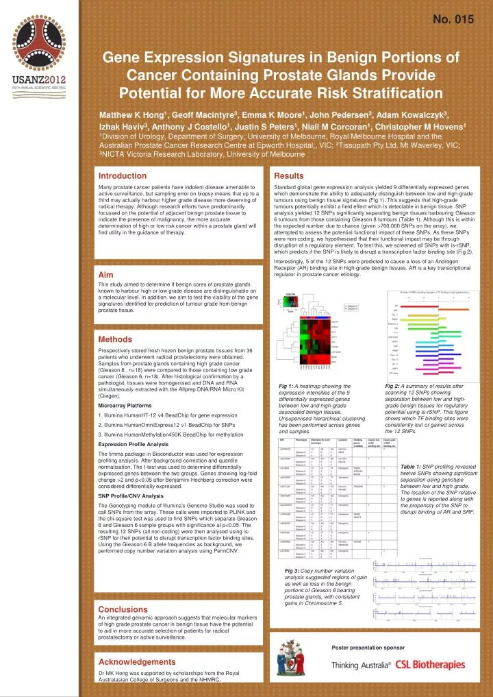
No. 015 Gene Expression Signatures in Benign Portions of Cancer Containing Prostate Glands Provide Potential for More Accurate Risk Stratification Matthew K Hong1, Geoff Macintyre3, Emma K Moore1, John Pedersen2, Adam Kowalczyk3, Izhak Haviv3, Anthony J Costello1, Justin S Peters1, Niall M Corcoran1, Christopher M Hovens1 1Division of Urology, Department of Surgery, University of Melbourne, Royal Melbourne Hospital and the Australian Prostate Cancer Research Centre at Epworth Hospital,, VIC; 2Tissupath Pty Ltd, Mt Waverley, VIC; 3NICTA Victoria Research Laboratory, University of Melbourne Results Standard global gene expression analysis yielded 9 differentially expressed genes which demonstrate the ability to adequately distinguish between low and high-grade tumours using benign tissue signatures (Fig 1). This suggests that high-grade tumours potentially exhibit a field effect which is detectable in benign tissue. SNP analysis yielded 12 SNPs significantly separating benign tissues harbouring Gleason 6 tumours from those containing Gleason 8 tumours (Table 1). Although this is within the expected number due to chance (given >700,000 SNPs on the array), we attempted to assess the potential functional impact of these SNPs. As these SNPs were non-coding, we hypothesised that their functional impact may be through disruption of a regulatory element. To test this, we screened all SNPs with is-rSNP, which predicts if the SNP is likely to disrupt a transcription factor binding site (Fig 2). Interestingly, 5 of the 12 SNPs were predicted to cause a loss of an Androgen Receptor (AR) binding site in high-grade benign tissues. AR is a key transcriptional regulator in prostate cancer etiology. Introduction Many prostate cancer patients have indolent disease amenable to active surveillance, but sampling error on biopsy means that up to a third may actually harbour higher grade disease more deserving of radical therapy. Although research efforts have predominantly focussed on the potential of adjacent benign prostate tissue to indicate the presence of malignancy, the more accurate determination of high or low risk cancer within a prostate gland will find utility in the guidance of therapy. Aim This study aimed to determine if benign cores of prostate glands known to harbour high or low grade disease are distinguishable on a molecular level. In addition, we aim to test the viability of the gene signatures identified for prediction of tumour grade from benign prostate tissue. Methods Prospectively stored fresh frozen benign prostate tissues from 36 patients who underwent radical prostatectomy were obtained. Samples from prostate glands containing high grade cancer (Gleason 8 , n=18) were compared to those containing low grade cancer (Gleason 6, n=18). After histological confirmation by a pathologist, tissues were homogenised and DNA and RNA simultaneously extracted with the Allprep DNA/RNA Micro Kit (Qiagen). Microarray Platforms 1. Illumina HumanHT-12 v4 BeadChip for gene expression 2. Illumina HumanOmniExpress12 v1 BeadChip for SNPs 3. Illumina HumanMethylation450K BeadChip for methylation Expression Profile Analysis The limma package in Bioconductor was used for expression profiling analysis. After background correction and quantile normalisation, The t-test was used to determine differentially expressed genes between the two groups. Genes showing log-fold change >2 and p<0.05 after Benjamini-Hochberg correction were considered differentially expressed. SNP Profile/CNV Analysis The Genotyping module of Illumina's Genome Studio was used to call SNPs from the array. These calls were imported to PLINK and the chi-square test was used to find SNPs which separate Gleason 8 and Gleason 6 sample groups with significance at p<0.05. The resulting 12 SNPs (all non-coding) were then analysed using is-rSNP for their potential to disrupt transcription factor binding sites. Using the Gleason 6 B allele frequencies as background, we performed copy number variation analysis using PennCNV. Fig 2: A summary of results after scanning 12 SNPs showing separation between low and high-grade benign tissues for regulatory potential using is-rSNP. This figure shows which TF binding sites were consistently lost or gained across the 12 SNPs. Fig 1: A heatmap showing the expression intensities of the 9 differentially expressed genes between low and high-grade associated benign tissues. Unsupervised hierarchical clustering has been performed across genes and samples. Table 1: SNP profiling revealed twelve SNPs showing significant separation using genotype between low and high grade. The location of the SNP relative to genes is reported along with the propensity of the SNP to disrupt binding of AR and SRF. Fig 3: Copy number variation analysis suggested regions of gain as well as loss in the benign portions of Gleason 8 bearing prostate glands, with consistent gains in Chromosome 5. Conclusions An integrated genomic approach suggests that molecular markers of high grade prostate cancer in benign tissue have the potential to aid in more accurate selection of patients for radical prostatectomy or active surveillance. Poster presentation sponsor Acknowledgements Dr MK Hong was supported by scholarships from the Royal Australasian College of Surgeons and the NHMRC.