Download
1 / 48
650 likes | 1.27k Vues
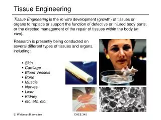
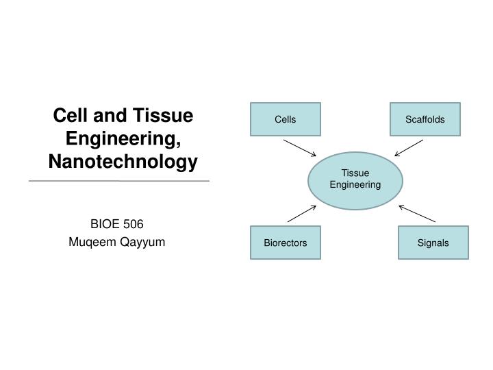
Cells. Scaffolds. Cell and Tissue Engineering, Nanotechnology. Tissue Engineering. BIOE 506 Muqeem Qayyum. Biorectors. Signals. Why?. In-vitro . In-vivo.

E N D
Cells Scaffolds Cell and Tissue Engineering, Nanotechnology Tissue Engineering BIOE 506 Muqeem Qayyum Biorectors Signals
Why? • In-vitro • In-vivo Because we can no longer to view a cell as self contained unit existing in a passive structural network. Thus to properly study the cell interactions it must be in a 3D environment. Image sourse: www.millipore.com/images farm1.static.flickr.com
EXTRACELLULAR MATRIX (ECM) • Consists of a 3D array of protein fibers and filaments embedded in a hydrated gel of glycosaminoglycans • ECM molecules • Glycosaminoglycans • Proteoglycans • Proteins Proteins in ECM act as either structural members or adhesion sites • Structural proteins • Collagen • Elastin • Adhesion proteins • Fibronectin • Laminin
Definitions • Tissue Engineering: • Interdisciplinary field addressing the improvement, repair, or replacement of tissue/organ function. • Nanotechnology: • Is study of gaining control of structures at the atomic, molecular levels. • Biocompatibility • Material should not elicit a significant or prolonged inflammatory response.4 • Biodegradability • The degraded products of the scaffold must have a safe route for removal from the host.4 • Mechanical Strength • The scaffold must be able to provide support to the forces applied to it and the surrounding tissues (especially true for the engineering of weight-bearing orthopedic tissues). 4
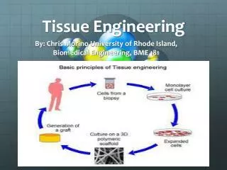
Tissue Engineering (TE) • Scaffolds Biomaterials, which may be natural or artificially derived, providing a platform for cell function, adhesion and transplantation • Cells Any class of cell, such as stem or mesenchymal cell • Signals Proteins and growth factors driving the cellular functions of interest • Bioreactor • System that supports a biologically active environment (ex. Cell culture) Image sourse: Stke.sciencemag.org, Nature.com
Barnes, C.P., Nanofiber technology: Designing the next generation of tissue engineering scaffolds1 Discussion
Nanofiber Technology • Techniques of nanofibers for tissue engineering • Characteristics of tissue engineering nanofibrous structures • Electrospinning of synthetic and natural polymers
3 techniques to achieve nanofibers for TE Self-assembly Phase separation Electrospinning
Self-assembly • Relies on non-covalent interactions to achieve spontaneously assembled 3D structure. • Biopolymers such as peptides and nucleic acids are used. Example is peptide-amphiphile (PA) • (A) Chemical structure of (PA) • (B) Molecular model of the PA showing the narrow hydrophobic tail to the bulkier peptide region • (C) Schematic of PA molecules into a cylindrical micelle.5 peptide-amphiphile Nanofiber
Phase separation • This process involves dissolving of a polymer in a solvent at a high temperature followed by a liquid–liquid or solid–liquid phase separation induced by lowering the solution temperature • Capable of wide range of geometry and dimensions include pits, islands, fibers, and irregular pore structures • Simpler than self-assembly a) powder, b) scaffolds with continuous network, c) foam with closed pores.4 SEM of nanofibrous scaffold with interconnected spherical macropores1
Electrospinning • This process involves the ejection of a charged polymer fluid onto an oppositely charged surface. • multiple polymers can be combined • control over fiber diameter and scaffold architecture A schematic of the electrospinning process to illustrate the basic phenomena and process components1
Overview Nanofiber No consensus of gold standard for creating native ECM
Scaffolds under Electrospinning • Goal of scaffolds • Properties • Materials • Synthetic • Natural • Limitations
Goals/Properties Goalof TE is to combine cell, scaffold (artificial ECM) and bioreactor to design and fabricate tissues and organs. Design scaffold with maximum control over: biocompatibility (chemical) biodegradability (mechanical) Scaffolds Properties • Biocompatible • Promote growth • Maintain 3-D structure • Non-immunogenic • Support tissue and cell forces
Synthetic Mimic mechanical properties (strength, elongation and knot retention) Mass production Degradation between hosts is minimal Natural Cell recognition Biodegradable Difficult degradation control between host Materials
Synthetic • Polyglycolic acid (PGA) • Highly crystalline, hydrophilic, byproduct is glycolic acid • Polylactic acid (PLA) • Hydrophobic, lower melting temperature, byproduct is lactic acid • Polydioxanone (PDO) • Highly crystalline • Polycaprolactone (PCL) • Semi-crystalline properties, easily co-polymerized, byproduct caproic acid • Blends • PGA-PLA • PGA-PCL • PLA-PCL • PDO-PCL
Poly(glycolic acid) (PGA) • Disadvantages: • fast degradation causes pH change • Tissue may require buffering capacity Advantages: • Biocompatible & biodegradable • bioabsorption (2-4 wks) • electrospinning yields diameters ~ 200 nm • Good choice for high strength and elasticity and fast degrading material SEM showing the random fiber arrangement (left) and the aligned fiber orientation (right) (1600× magnification).
PGA • Spinning orientation affects scaffold elastic modulus • The overall results exhibit a correlation between the fiber diameter and orientation and the elastic modulus and strain to failure Fig. summarizes mechanical properties in terms of the elastic modulus and strain at failure of PGA in a uniaxial model.
Poly(lactic acid) (PLA) • Disadvantages: • Larger diameter fibers ~ microscale Advantages: • Biocompatible & biodegradable • bioabsorption (30 wks) • Good choice for drug delivery do to predictable degradation SEM showing the random oriented PLA from cholorform (left) and the randomly oriented PLA from HFP (right)
PGA + PLA blends (PLGA) • Group found: • Hydrophilicity proportional to composition of copolymer • Degradation rate proportional to composition of copolymer Group Tested copolymers of following ratios: • 75%PLA–25%PGA • 50%PLA–50%PGA, • blended PLA and PGA together in HFP at ratios of 100:0, 75:25, 50:50, 25:75
PLGA • Applications of PLGA: • cardiac tissue in mice for tissue regeneration • individual cardiomyocytes attachment at seeding • scaffold loaded with antibiotics for wound healing mechanical properties, such as tangential modulus, peak stress, and strain to failure, of these copolymers and blends appear to be controlled by the fiber/polymer composition
Advantages: Biocompatible & biodegradable degradation rate between PGA/PLA Shape memory Excellent flexibility Modulus comparable collagen and elastin Good source for future vascular grafts Polydioxanone (PDO) • Disadvantages: • Lack on knot retention • Lack of adaptability to developing tissue SEM of 180 nm diameter randomly oriented fibrous structures Results of the fiber diameter analysis versus PDO concentration illustrating the linear relationship between electrospinning solution concentration and fiber diameter.
Polycaprolactone (PCL) • Disadvantages: • No shape retention (highly elastic) Advantages: • Biocompatible & biodegradable • Inexpensive • Highly elastic • Slow degradation (1 – 2 yrs) • Good choice for Human mesenchymal stem cells (hMSCs) seeding to induce differentiation • Applications: • Bone tissue strengthening • Cardiac grafts • Collagen and cellular interaction • Differentiation with MSC cells
Advantages: PGA – high stress tolerance PCL – highly elastic Optimum combination PCL/PGA ~ 1/3 Longer degradation time ~ 3 months (PCL-2 yrs, PGA 2-4 wks) PGA + PCL blend
PLA + PCL blends • Disadvantages: • Decreasing PLA+PCL ratios decreases strain capacity, optimized at 95:5 Advantages: • Greater elasticity than PGA+PCL • Similar tensile strength to PLA • ~5% addition of PCL increased strain by 8 fold • Overall best synthetic ECM for cardiac applications Strain to failure of a tissue matrix a function of both composition (varying blend ratios of PLA and PCL) and fiber alignment
PDO + PCL blends Advantages: • PCL high elasticity • PDO shape memory • Disadvantages: • lower tensile capacity than PDO • lower elasticity than PDO • larger fiber diameter Peak stress values for various PCL concentrations versus PDO:PCL ratio and orientation of the testing specimen * Further investigation needed Strain at break values for various PCL concentrations versus PDO:PCL ratio and orientation of the testing specimen
Natural • Elastin • Gelatin collagen • Fibrillar collagen • Collagen blends • Fibrinogen
Elastin • Advantages: • Linearly elastic biosolid • Insoluble and hydrophobic • Critical role in shape and energy recovery for organs • Disadvantages: • Less elastic than native elastin • Needs to be combined with PDO to increase tensile strength • Fiber ~300 nm (not as small as PDO ~ 180 nm) • Varying diameter SEM of electrospun elastin scaffold at 250 mg/ml.
Collagen • Gelatin (denatured collagen) • Advantages: • Biocamptibale and biodegradable • Inexpensive • Disadvantages: • Quick to dissolve • Fibril-forming (Types I, II, III) • Most abundant natural polymers in body • Important role in ECM • Type I: principal structure in ECM • Type II: pore size and fiber diameter easily controlled • Type III: still under investigation
Collagen blends (1st attempts) • Artery • Intima: innermost layer, composed of single layer of endothelial cells on basement membrane of elastin and Type IV collagen • Media: thickest layer, several layers consisting of collagen type I & III, elastin and proteoglycans • Adventitia: made of fibroblast and collagen type I (Left) Photograph of 2 and 4 mm ID electrospun scaffolds. (Middle) SEM of tubular electrospun composite. (Right) SEM of electrospun 40:40:20 blend of collagen type I, collagen type III, and elastin with random orientation.
Collagen blends (1st attempts) • Collagen Type I & III + PDO • indication that blends of PDO and collagen may match mechanical and morphological requirements of a blood vessel's microenvironment (similar to PDO section). Tangential modulus presented as a function of the ratio of PDO to collagen type I & III. collagen I highest tensile capacity, optimal ratio for all collagens was 70:30 collagen-PDO.
Globular proteins • Fibrinogen (protein in blood plasma – wound healing) • Low concentration produced fibers within range of fibrinogen fibers in plasma clots (80, 310, 700 nm) • High surface area to volume ratio: increases area available for clot formation • Stress capacity comparable to collagen (80-100 MPa) the linear relationship between concentration and fiber diameter composing the structures produced.
Globular proteins • Hemoglobin & myoglobin • Fiber sizes 2-3 µm & 490 – 990 nm • Spun with fibrinogen for clotting and healing improvements • High porosity means higher oxygenation • Clinical applications: • Drug delivery • Hemostatic bandages • Blood substitutes SEM of electrospun hemoglobin in 2,2,2-Trifluoroethanol at 150 mg/ml (A), 175 mg/ml (B), and 200 mg/ml
Summary Scaffolds: • Electrospinning viable for both synthetic and biological scaffolds/mats • synthetic polymers • PGA, PLA and PLGA most commonly used • PDO most similar to Elastin collagen blend (limited by shape memory) • PCL most elastic and mixed frequenlty with other material • Provide nanoscale physical features • Natural polymers • Collagen Type I & III + PDO: best possible match for blood vessels Limitations on Scaffolds • Mechanical material failure • Immunogenic reaction to material
Dalby, M.J., The control of human mesenchymal cell differentiation using nanoscale symmetry and disorder2 Discussion
Overview 3D Scaffolds electron beam lithography (EBL) Image source: 6
TE for Bone Mesenchymal: Derived from mesoderm tissue consists of undifferentiated cells loosely organized within ECM.6 Osteoprogenitor cells: located in periosteum and bone marrow giving rise to osteoblasts. Electron-beam lithography is a technique that employs a focused beam of electrons in order to pattern a mask or a silicon wafer. Image source: 6
Experiment Experiment looking to model osteoblastic differentiation of two cell types, osteoprogenitors and MSCs, and their interactions following culture on nanofeatures of different symmetry and with varying degrees of disorder.
Results (osteoprogenitors) • Immunofluorescence was used to detect expression of the bone-specific ECM proteins osteopontin (OPN) and osteocalcin (OCN) by osteoprogenitors in response to the materials. • good cell density • cells remained fibroblastic appearance • lack of OPN & OCN production • decerase osteoprogenitors • cell density • poor cell adhesion SQ HEX
Results (2) • formation of dense aggregates • raised levels of OPN and OCN DSQ50 • more-dense cell growth but • only limited OPN or OCN levels RAND
Results (MSCs) • Two MSC batches were cultured: one for 21 days before labeling OPN and OCN and a second population for 28 days before alizarin red staining (stain for calcium present in bone mineral, indication of osteospecific differentiation). • fibroblastic in appearance • highly elongated and aligned morphology • more typical osteoblastic • morphology • negligible OPN or OCN • presence SQ RAND
Results (2) • typical osteoblastic morphology • expressed foci of OPN • lack of OCN expression DSQ20 • discrete areas of intense • cell aggregation • early nodule formation • positive regions of OPN & OCN 28 days allowed positive identification of mature bone nodules noted. DSQ50
Osteospecific macroarrays • To compare MSC differentiation for cells cultured on (1) untreated cells cultured on a planar control, (2) planar material with dexamethasone (DEX) (steroid to induce bone formation) and (3) DSQ50 Cells cultured with DEX exhibited the highest levels of osteogenic up-regulation (24 gene hits) followed by MSCs cultured on DSQ50 (11 gene hits). Cells cultured on the planar control, however, demonstrated only three gene hits.
qPCR • To confirm these results, a quantitative polymerase chain reaction (qPCR) was used with primers for the bone marker genes OCN and alkaline phosphatase In agreement with the macroarray data, MSCs cultured with DEX or on the DSQ50 material had significantly higher expressions of genes of interest.
Summary • The qPCR and macroarray results together clearly indicated that the disordered materials have an osteogenic potential close to that of DEX and a higher osteogenic potential than that of highly ordered or totally random topographies. • It was shown that surface topography caused significant changes in MSC response and that topography may induce a broader response than simply changing osteoblast-specific genes.
Summary (2) • Results demonstrate that highly ordered nanotopographies produce low to negligible cellular adhesion and osteoblastic differentiation. • Cells on random nanotopographies exhibited a more osteoblastic morphology by 14 days • It seems more likely that as long as cell spreading is permitted by the surface, cells will follow a classical differentiation timelines.
Reference • Barnes, C.P., Nanofiber technology: Designing the next generation of tissue engineering scaffolds. • Dalby, M.J., The control of human mesenchymal cell differentiation using nanoscale symmetry and disorder. • Gonsalves, K.E., Biomedical Nanostructures, Wiley 2008. • Laurencin, C.T., Nanotechnology and Tissue Engineering: The Scaffold, CRC Press. 2008 • Hartgerink, J.D., Self-assembly and Mineralization of Peptide-amphiphile Nanofibers, V. 294, No. 5547. 2001. • Novakovic, G., Culture of Cells for Tissue Engineering. Wiley, 2006