Download
1 / 13
130 likes | 343 Vues
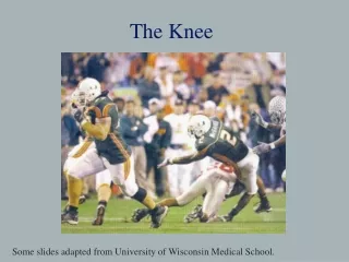
The KNEE. Willis McGahee injury –2003 Fiesta Bowl. Napolean Kaufman. This injury is called an “ unhappy TRIAD ” and we’ll look at it more closely later in the period. Knee Articulations (Sketch the bones that make up the knee joint). -the knee consists of 4 different bones. Femur. Patella.

E N D
Willis McGahee injury –2003 Fiesta Bowl Napolean Kaufman This injury is called an “unhappy TRIAD” and we’ll look at it more closely later in the period.
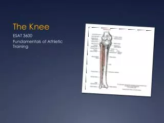
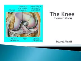
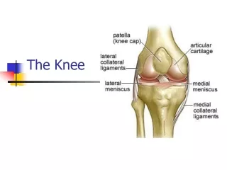
Knee Articulations(Sketch the bones that make up the knee joint) -the knee consists of 4 different bones Femur Patella Tibia Fibula -hinge joint with a slight pivot (extension, flexion and medial/lateral rotation) bathed in a synovial fluid found in the joint capsule that surrounds the joint. -largest and most complex joint in the body
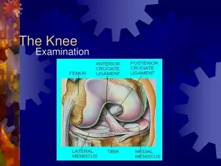
What view? How is the knee held together? Anterior -the knee is so complex because of the interplay of the ligaments and accessory parts Patellar tendon Anterior cruciate ligament Posterior cruciate ligament Articular cartilage Medial collateral ligament Lateral miniscus Lateral collateral ligament Medial miniscus
A closer look at the ligaments Medial collateral ligament (MCL): stretches from medial epicondyle of the femur to the medial tibial condyle; protects medial side of knee from being opened by a lateral force VALGUS Lateral collateral ligament (LCL): lateral epicondyle of the femur to the head of the fibula; protects lateral side from a medial force VARUS Anterior cruciate ligament (ACL): lateral condyle of femur to the anterior intercondylar area; limits ability of tibia to be pushed anterior too far relative to the femur. Cross = crucitate Posterior cruciate ligament (PCL): medial condyle of the femur to the posterior intercondylar area; prevents posterior movement of the tibia relative to the femur Lateral and Medial minisci: articular disks of cartilage; protect the ends of the bones from rubbing on each other, provide shock absorption -video
Well what happens when some of these forces go beyond what your knee can handle? The Unhappy TRIAD Shaun Livington -video The menisci can be torn/cracked when forcefully rotated or excessively bent The ACL can be torn by a twisting or bending motion The MCL can be torn by a lateral force
The Unhappy TRIAD “…a common combination of three different injuries to the knee: ACL, MCL and meniscus tear.” Results from a PLANT and TURN movement coupled with a LATERAL BLOW -commonly found in contact or high impact sports Let’s look at each individual injury a little more closely. . . MCL Tear -ligament provides stability -least problematic of the three -often MCL will regrow on its own or you can live without one -produce pain and swelling, some instability and decreased range of motion
Torn Meniscus -help provide cushion and reduce friction and stresses on the bones -treated surgically due to slow healing process if left alone -KNEE SCOPE -common along with ACL tears -if untreated produces pain, swelling, and may catch on movement Video: http://www.youtube.com/watch?v=EC6J_pj0SRQ Torn ACL -worst and most significant injury of the triad -knee loses stability, motion and may “buckle” under regular walking -surgery is the only option -will take months to a year for full recovery Video: http://www.youtube.com/watch?v=q96M0jRqn7k&NR=1 Video: http://www.youtube.com/watch?v=i8EpT3uCVWU
Steps to ACL Reconstruction Surgery 1. Knee Arthroscopy -setting up of camera and tools through 2 holes (portals) -the doctor will explore the knee to see the amounts of damage to the triad areas 2. Meniscectomy -cleaning up of any debris (torn meniscus, smooth articular cartilage) in the knee along with removal of torn ACL -increase in size of femoral notch (for new ACL graft) 3. Graft Harvest -graft is taken from patellar tendon or hamstrings -graft is stretch and prepped to be inserted 4. Graft -graft is passed through holes drilled into tibia and femur and screwed into place 5. The incisions are closed, dressings applied and recovery begins
Rehabilitation It is very important that rehabilitation is started the moment the patient wakes up after surgery Phase I (0-2 weeks)–regaining range of motion and minimizing swelling -ice and elevated, while staying off feet (crutches) -end of phase: knee full extension 90 degrees flexion Phase II (2-6 weeks) –maintain full extension and increase flexion -daily exercises to bring back quad strength -end of phase: nearly full range of motion; increase exercise intensity monitored by post exercise swelling Phase III ( > 6 weeks) –building back strength; allowing ligament to heal -closed-chain kinetic exercises: foot planted on floor unable to move (ex squats) • 3 months till start agility exercises • Difficult to set timeline for return to sport person to person BRACE