Download
1 / 61
620 likes | 951 Vues
Inflammatory Disorders of the Skin. Objectives.

E N D
Objectives 1. Describe and discuss, Dermatitis, Acne Vulgaris , Urticaria, Psoriasis, Seborrheic Keratosis. Scleroderma and Systemic Lupus Erythematous as to definition, etiology, pathophysiology, signs and symptoms, diagnosis, medical and nursing management. 2. Apply the nursing process for clients with inflammatory disorders of the skin. 3. Recognize systemic disorders with dermatologic symptoms.
Content 1. Dermatitis a. Etiology b. Pathophysiology c. Signs and Symptoms d. Diagnostic findings e. Medical management f. Nursing management
Content 2. Acne Vulgaris a. Etiology b. Pathophysiology c. Signs and Symptoms d. Diagnostic findings e. Medical management f. Nursing management
Content 3. Urticaria a. Etiology b. Pathophysiology c. Signs and Symptoms d. Diagnostic findings e. Medical management f. Nursing management
Content 4. Psoriasis a. Etiology b. Pathophysiology c. Signs and Symptoms d. Diagnostic findings e. Medical management
Content 5. Seborrheic Keratosis a. Etiology b. Pathophysiology c. Signs and Symptoms d. Diagnostic findings e. Medical management f. Nursing management
Content 6. Scleroderma a. Etiology b. Pathophysiology c. Signs and Symptoms d. Diagnostic findings e. Medical management f. Nursing management
Content 7. Systemic Lupus Erythematous a. Etiology b. Pathophysiology c. Signs and Symptoms d. Diagnostic findings e. Medical management f. Nursing management
Content 8. Nursing Process a. Assessment b. Nursing Diagnosis c. Planning d. Implementation e. Evaluation
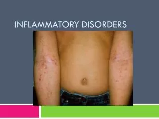
Dermatitis • A general term used to describe inflammation of the skin. Description • Most types of dermatitis are characterized by an itchy pink or red rash. • Contact dermatitis is an allergic reaction to something that irritates the skin and is manifested by one or more lines of red, swollen, blistered skin that may itch or seep. • It usually appears within 48 hours after touching or brushing against a substance to which the skin is sensitive. • The condition is more common in adults than in children.
Dermatitis Contact dermatitis of the (left) face and (right) wrist
Contact dermatitis • Can occur on any part of the body, but it usually affects the hands, feet, and groin. • Contact dermatitis usually does not spread from one person to another, nor does it spread beyond the area exposed to the irritant unless affected skin comes into contact with another part of the body.
Contact dermatitis • However, in the case of some irritants, such as poison ivy, contact dermatitis can be passed to another person or to another part of the body.
Atopic Dermatitis • This form of dermatitis, commonly referred to as eczema, is a chronic condition that causes itchy, inflamed skin. • Most often, it occurs in the folds of the elbows, backs of the knees or the front of the neck. • It tends to flare periodically and then subside for a time, even up to several years. • The exact cause of this skin disorder is unknown, but it may result from a malfunction in the body's immune system.
Dermatitis • Pathophysiology and Etiology • Types: Allergic contact; primary irritant • Assessment Findings • Blood vessel dilation; itching; vesiculation • Skin patch test; visual examination
Dermatitis • Medical Management • Flushing skin with cool water; topical lotions; wet dressings; corticosteroids • Nursing Management • Client teaching: Avoid contact with soap, topical substances; preserve skin integrity
Acne Vulgaris Acne of (left) the face and (right) the chest
Acne Vulgaris • Pathophysiology and Etiology • Overproduction of sebum • Assessment Findings • Comedones (blackhead); oily scalp • Visual examination • Medical Management • Gentle facial cleansing; drying agents containing benzoyl peroxide • Topical and oral drugs and antibiotics • Removal with instruments
Acne • Develops as a result of blockages in follicles. Hyperkeratinization and formation of a plug of keratin and sebum (a microcomedo) is the earliest change. • Enlargement of sebaceous glands and an increase in sebum production occur with increased androgen production . • The microcomedo may enlarge to form an open comedo (blackhead) or closed comedo (whitehead). • Increased sebum production provides an environment for the overgrowth of Propionibacterium acnes.
Acne Vulgaris • Surgical Management • Dermabrasion • Nursing Management • Client teaching • Cleanliness: Face and hair • Avoid cosmetics, Manipulation of lesions • Precautions for pregnant women: Risk associated with systemic oral isotretinoin
Urticaria • A vascular reaction pattern of the skin marked by the transient appearance of smooth, slightly elevated patches that are more red or more pale than the surrounding skin and are accompanied by severe itching. • Also called hives.
Non-allergic urticaria • Mechanisms other than allergen-antibody interactions are known to cause histamine release from mast cells. For instance, a diverse group of signaling substances called neuropeptides have been found to be involved in emotionally induced urticaria.
Urticaria • An acute or chronic condition characterized by the appearance of itchy weals on the skin. • The cause may be an allergy to certain foods , drugs, emotional stress, or local skin irritation resulting from contact with certain plants. • Athletes sometimes develop hives while exercising (exercise-induced urticaria). The hives are small and seem to develop in response to the release of histamines associated with the increase in body temperature produced by exercise.
Treatment & Management • Most treatment plans for urticaria involve being aware of one's triggers. • If one's triggers can be identified then outbreaks can often be managed by limiting one's exposure to these situations.
Drug treatment • Typically in the form of Antihistamines such as diphenhydramine, hydroxyzine, cetirizine and other H1 receptor antagonists. These are taken on a regular basis to protective effect, lessening or halting attacks. • For some people, H2-receptor antagonists such as cimetidine (Tagamet) and ranitidine (Zantac) can also help control symptoms either protectively or by lessening symptoms when an attack occurs. • When taken in combination with a H1 antagonist it has been shown to have a synergistic effect which is more effective than either treatment alone.
Psoriasis • Pathophysiology and Etiology • Likely genetic predisposition • Keratinocytes; plaque • Assessment Findings • Erythema with silvery scales; lesions • Visual examination; skin biopsy • Medical Management • Symptomatic treatment • Drug therapy; biologic therapy (immunotherapy - interferon) • Photochemotherapy (fiberoptic probe)
Psoriasis • Named for the Greek word psōra meaning "itch," psoriasis is a chronic, non-contagious disease characterized by inflamed lesions covered with silvery-white scabs of dead skin.
Pathophysiology • Normal skin cells mature and replace dead skin every 28–30 days. • Psoriasis causes skin cells to mature in less than a week. • Because the body can't shed old skin as rapidly as new cells are rising to the surface, raised patches of dead skin develop on the arms, back, chest, elbows, legs, nails, folds between the buttocks, and scalp.
Psoriasis is considered mild if it affects less than 5% of the surface of the body; moderate, if 5–30% of the skin is involved, and severe, if the disease affects more than 30% of the body surface.
Nursing Process: The Client With Psoriasis • Assessment • Skin integrity; appearance • Family history of psoriasis • Triggering factors • Diagnosis, Planning, and Interventions • Impaired skin integrity • Disturbed body image
Nursing Process: The Client With Psoriasis • Evaluation of Expected Outcomes • Improved integrity and appearance of skin • Reduced itching; copes effectively with altered appearance
Seborrheic Keratosis A superficial, benign, verrucose lesion consisting of proliferating epidermal cells enclosing horn cysts, usually appearing on the face, trunk, or extremities in adulthood.
Sign And Symptoms • The growths resemble flattened or raised warts, but have no viral origins and may exhibit a variety of colors, from pink or yellow through brown and black. • Because only the top layers of the epidermis are involved, seborrheic keratoses are often described as having a "pasted-on" appearance.
Etiology • A mutation of a gene coding for a growth factor receptor (FGFR3), has been associated with seborrheic keratosis.
Treatment • Because the tumors are rarely painful, treatment is not often necessary. • If a growth becomes excessively itchy, or if it is irritated by clothing or jewelry, cryosurgery has been found to be highly effective in their removal. • With resemblance to malignant melanomas, which has sometimes led to a misdiagnosis of the cancerous lesions. If there is any doubt, a skin biopsy will allow a physician to make a correct diagnosis.
Scleroderma • Scleroderma is a progressive disease that affects the skin and connective tissue (including cartilage, bone, fat, and the tissue that supports the nerves and blood vessels throughout the body). • There are two major forms of the disorder. Localized scleroderma mainly affects the skin. Systemic scleroderma, which is also called systemic sclerosis, affects the smaller blood vessels and internal organs of the body.
Scleroderma • Is an autoimmune disorder, which means that the body's immune system turns against itself. In scleroderma, there is an overproduction of abnormal collagen (a type of protein fiber present in connective tissue). This collagen accumulates throughout the body, causing hardening (sclerosis), scarring (fibrosis), and other damage.
Therapy • There is no cure for every patient with scleroderma, though there is treatment for some of the symptoms, including drugs that soften the skin and reduce inflammation. Some patients may benefit from exposure to heat. • A range of NSAIDs (nonsteroidal anti-inflammatory drugs) can be used to ease symptoms, such as naproxen. If there is esophageal dysmotility .Care must be taken with NSAIDs as they are gastric irritants, and so a proton pump inhibitor (PPI) such as omeprazole can be given in conjunction.
Treatment • Immunosuppressant drugs, such as mycophenolate mofetil (Cellcept®) or cyclophosphamide are sometimes used to slow the progress. • Digital ulcerations and pulmonary hypertension can be helped by prostacyclin (iloprost) infusion. Iloprost increases blood flow by relaxing the arterial wall.
Sytemic Lupus Erythematous • Lupus is a condition characterized by chronic inflammation of body tissues caused by autoimmune disease. • Autoimmune diseases are illnesses that occur when the body's tissues are attacked by its own immune system.