Download
1 / 37
380 likes | 523 Vues
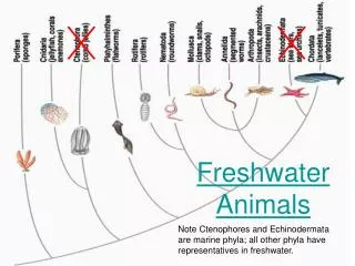
Freshwater animals show adaptations that reduce water uptake and conserve solutes Desert and marine animals face desiccating environments that can quickly deplete body water Osmoregulation regulates solute concentrations and balances the gain and loss of water

E N D
Freshwater animals show adaptations that reduce water uptake and conserve solutes • Desert and marine animals face desiccating environments that can quickly deplete body water • Osmoregulation regulates solute concentrations and balances the gain and loss of water • Excretion gets rid of metabolic wastes
Osmoregulation balances the uptake and loss of water and solutes • Osmoregulation is based largely on controlled movement of solutes between internal fluids and the external environment • Osmoregulators expend energy to control water uptake and loss in a hyperosmotic or hypoosmotic environment
Gain of water and salt ions from food and by drinking seawater Osmotic water loss through gills and other parts of body surface Excretion of salt ions and small amounts of water in scanty urine from kidneys Excretion of salt ions from gills Osmoregulation in a saltwater fish
Osmotic water gain through gills and other parts of body surface Uptake of water and some ions in food Uptake of salt ions by gills Excretion of large amounts of water in dilute urine from kidneys Osmoregulation in a freshwater fish
Transport Epithelia • Transport epithelia are specialized cells that regulate solute movement • They are essential components of osmotic regulation and metabolic waste disposal • They are arranged in complex tubular networks • An example is in salt glands of marine birds, which remove excess sodium chloride from the blood
Nasal salt gland Nostril with salt secretions
Lumen of secretory tubule Vein Capillary Artery Secretory tubule NaCl Transport epithelium Direction of salt movement Blood flow Secretory cell of transport epithelium Central duct
Nucleic acids Proteins Amino acids Nitrogenous bases —NH2 Amino groups Most aquatic animals, including most bony fishes Mammals, most amphibians, sharks, some bony fishes Many reptiles (including birds), insects, land snails Ammonia Urea Uric acid
Ammonia • Animals that excrete nitrogenous wastes as ammonia need lots of water • They release ammonia across the whole body surface or through gills
Urea • The liver of mammals and most adult amphibians converts ammonia to less toxic urea • The circulatory system carries urea to the kidneys, where it is excreted
Uric Acid • Insects, land snails, and many reptiles, including birds, mainly excrete uric acid • Uric acid is largely insoluble in water and can be secreted as a paste with little water loss
Excretory Processes • Most excretory systems produce urine by refining a filtrate derived from body fluids • Key functions of most excretory systems: • Filtration: pressure-filtering of body fluids • Reabsorption: reclaiming valuable solutes • Secretion: adding toxins and other solutes from the body fluids to the filtrate • Excretion: removing the filtrate from the system
Capillary Filtration Excretory tubule Filtrate Reabsorption Secretion Urine Excretion
Protonephridia: Flame-Bulb Systems • A protonephridium is a network of dead-end tubules lacking internal openings • The smallest branches of the network are capped by a cellular unit called a flame bulb • These tubules excrete a dilute fluid and function in osmoregulation
Nucleus of cap cell Cilia Interstitial fluid filters through membrane where cap cell and tubule cell interdigitate (interlock) Tubule cell Flame bulb Protonephridia (tubules) Tubule Nephridiopore in body wall
Metanephridia • Each segment of an earthworm has a pair of open-ended metanephridia • Metanephridia consist of tubules that collect coelomic fluid and produce dilute urine for excretion
Coelom Capillary network Bladder Collecting tubule Nephridio- pore Nephrostome Metanephridium
Malpighian Tubules • In insects and other terrestrial arthropods, Malpighian tubules remove nitrogenous wastes from hemolymph and function in osmoregulation • Insects produce a relatively dry waste matter, an important adaptation to terrestrial life
Digestive tract Rectum Hindgut Intestine Midgut (stomach) Malpighian tubules Anus Feces and urine Salt, water, and nitrogenous wastes Malpighian tubule Rectum Reabsorption of H2O, ions, and valuable organic molecules HEMOLYMPH
Nephrons and associated blood vessels are the functional unit of the mammalian kidney • Kidneys, the excretory organs of vertebrates, function in both excretion and osmoregulation • The mammalian excretory system centers on paired kidneys, which are also the principal site of water balance and salt regulation • Each kidney is supplied with blood by a renal artery and drained by a renal vein • Urine exits each kidney through a duct called the ureter • Both ureters drain into a common urinary bladder
Posterior vena cava Renal artery and vein Kidney Renal medulla Aorta Renal cortex Ureter Renal pelvis Urinary bladder Urethra Ureter Excretory organs and major associated blood vessels Section of kidney from a rat Kidney structure Juxta- medullary nephron Cortical nephron Afferent arteriole from renal artery Glomerulus Bowman’s capsule Proximal tubule Peritubular capillaries Renal cortex Collecting duct SEM 20 µm Efferent arteriole from glomerulus Renal medulla Distal tubule To renal pelvis Collecting duct Branch of renal vein Descending limb Loop of Henle Nephron Ascending limb Vasa recta Filtrate and blood flow
Structure and Function of the Nephron and Associated Structures • The mammalian kidney has two distinct regions: an outer renal cortex and an inner renal medulla • The nephron, the functional unit of the vertebrate kidney, consists of a single long tubule and a ball of capillaries called the glomerulus
Filtration of the Blood • Filtration occurs as blood pressure forces fluid from the blood in the glomerulus into the lumen of Bowman’s capsule • Filtration of small molecules is nonselective • The filtrate in Bowman’s capsule mirrors the concentration of solutes in blood plasma
Pathway of the Filtrate • From Bowman’s capsule, the filtrate passes through three regions of the nephron: the proximal tubule, the loop of Henle, and the distal tubule • Fluid from several nephrons flows into a collecting duct
Blood Vessels Associated with the Nephrons • Each nephron is supplied with blood by an afferent arteriole, a branch of the renal artery that divides into the capillaries • The capillaries converge as they leave the glomerulus, forming an efferent arteriole • The vessels divide again, forming the peritubular capillaries, which surround the proximal and distal tubules
From Blood Filtrate to Urine: A Closer Look • Filtrate becomes urine as it flows through the mammalian nephron and collecting duct • Secretion and reabsorption in the proximal tubule greatly alter the filtrate’s volume and composition • Reabsorption of water continues as filtrate moves into the descending limb of the loop of Henle
In the ascending limb of the loop of Henle, salt diffuses from the permeable tubule into the interstitial fluid • The distal tubule regulates the K+ and NaCl concentrations of body fluids • The collecting duct carries filtrate through the medulla to the renal pelvis and reabsorbs NaCl • The mammalian kidney conserves water by producing urine that is much more concentrated than body fluids
Proximal tubule Distal tubule Nutrients NaCl H2O HCO3– K+ HCO3– H2O NaCl H+ H+ NH3 K+ CORTEX Descending limb of loop of Henle Thick segment of ascending limb Filtrate H2O Salts (NaCl and others) HCO3– H+ Urea Glucose; amino acids Some drugs NaCl H2O OUTER MEDULLA NaCl Thin segment of ascending limb Collecting duct Key Urea NaCl Active transport Passive transport H2O INNER MEDULLA
Solute Gradients and Water Conservation • The cooperative action and precise arrangement of the loops of Henle and collecting ducts are largely responsible for the osmotic gradient that concentrates the urine • NaCl and urea contribute to the osmolarity of the interstitial fluid, which causes reabsorption of water in the kidney and concentrates the urine
Osmolarity of interstitial fluid (mosm/L) 300 300 100 300 100 300 300 H2O NaCl H2O CORTEX Active transport 200 400 400 400 H2O NaCl H2O Passive transport NaCl H2O H2O OUTER MEDULLA H2O NaCl H2O 400 600 600 600 H2O NaCl H2O Urea H2O NaCl H2O 700 900 900 Urea H2O H2O NaCl INNER MEDULLA Urea 1200 1200 1200
The countercurrent multiplier system involving the loop of Henle maintains a high salt concentration in the kidney • This enables the kidney to form concentrated urine
The collecting duct conducts filtrate through the osmolarity gradient, and more water exits the filtrate by osmosis • Urea diffuses out of the collecting duct as it traverses the inner medulla • Urea and NaCl form the osmotic gradient that enables the kidney to produce urine that is hyperosmotic to the blood
Regulation of Kidney Function • The osmolarity of the urine is regulated by nervous and hormonal control of water and salt reabsorption in the kidneys • Antidiuretic hormone (ADH) increases water reabsorption in the distal tubules and collecting ducts of the kidney
Osmoreceptors in hypothalamus Thirst Hypothalamus Drinking reduces blood osmolarity to set point ADH Increased permeability Pituitary gland Distal tubule H2O reab- sorption helps prevent further osmolarity increase STIMULUS The release of ADH is triggered when osmo- receptor cells in the hypothalamus detect an increase in the osmolarity of the blood Collecting duct Homeostasis: Blood osmolarity
The renin-angiotensin-aldosterone system (RAAS) is part of a complex feedback circuit that functions in homeostasis
Homeostasis: Blood pressure, volume Increased Na+ and H2O reab- sorption in distal tubules STIMULUS: The juxtaglomerular apparatus (JGA) responds to low blood volume or blood pressure (such as due to dehydration or loss of blood) Aldosterone Arteriole constriction Adrenal gland Angiotensin II Distal tubule Angiotensinogen JGA Renin production Renin
Animations and Videos • Water Uptake • The Human Kidney • Bozeman – Osmoregulation • The Mammalian Kidney • Chapter Quiz Questions – 1 • Chapter Quiz Questions – 2