Download
1 / 16
170 likes | 568 Vues
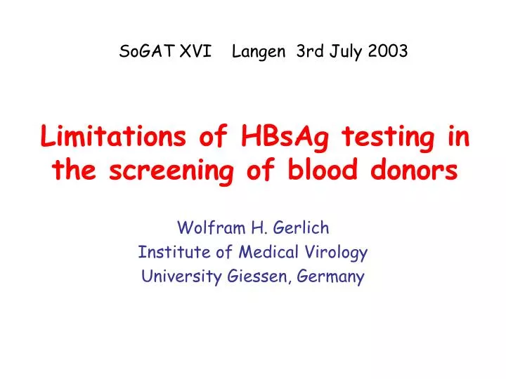
SoGAT XVI Langen 3rd July 2003 . Limitations of HBsAg testing in the screening of blood donors. Wolfram H. Gerlich Institute of Medical Virology University Giessen, Germany. Problems of HBsAg tests. Detection limit difficult to define Unclear HBsAg unit WHO unit not often applied

E N D
SoGAT XVI Langen 3rd July 2003 Limitations of HBsAg testing in the screening of blood donors Wolfram H. Gerlich Institute of Medical Virology University Giessen, Germany
Problems of HBsAg tests • Detection limit difficult to define • Unclear HBsAg unit • WHO unit not often applied • new standard preparation in planning • Paul Ehrlich Inst. (PEI) unit since 1975 • Standard and units changed over the years • Nanogram units of test producers • too high and discordant • Old PEI unit = 1 ng active HBsAg = 2 WHO unit • HBV genotypes react differently • Different HBsAg preparations react differently
Anti-a antiserum raised with subsequent injections of genotypes A, D, and C
Problems of HBsAg tests • HBV genotypes react differently • WHO provides only standard for genotype A • Worldwide are genotypes B,C und D predominant • Genotype F was not detected for years with some tests • Insufficient sensitivity • Best possible ELISA detects 10pg HBsAg/ml • Or 106 HBV particles/mL • if excessive HBsAg is not present in very early phase • HBsAg is highly variable under immune selection • after post exposure immunisation • during reactivation • Consequence: false negative results
Problems of HBsAg tests • HBV genotypes react differently • WHO standard only genotype A • Worldwide genotypes B,C und D predominant • Genotype F not detected for years with some tests • Insufficient sensitivity even for genotype A • Best possible ELISA detects 10pg HBsAg/ml • This corresponds to 106 HBV particles/mL • if excessive HBsAg is not present in early phase • HBsAg is highly variable under immune selection • after post exposure immunisation • during reactivation • Consequence: false negative results
HBsAg-negative, HBV-infectious blood donation : the donor • 16th July 2002 • HBsAg in AxSym negative • 17th September 2002 • AxSym (Abbott) positive s/co=2.4 • Enzygnost (Dade Behring) negative s/co=0.8 • 21st September 2002 • Enzygnost positive s/co=1.9 • HBV-DNA positive3200 ge/mL • Anti-HBc total antibody and IgM and HBeAg negative Meisel et al, submitted
HBsAg-negative, HBV-infectious blood donation: the recipient • 22. July 2002 • receives erythrocyte concentrate from the pre-seroconversion donation • HBsAg and anti-HBc negative • 19. November 2002 • HBsAg positive, HBV DNA 1.8x10¹⁰ genomes/mL • No hepatitis, develops persistent viremia • anti-HBc still negative, later positive • HBV DNA sequence of donor and recipient • identical, genotype A, in Germany predominant variant • S-gene without mutations
HBsAg-negative, HBV-infectious blood donation : the donor • 16th July 2002 • HBsAg in AxSym negative • HBV DNA ca 2000 genomes/mL • 17th September 2002 • AxSym (Abbott) positive s/co=2.4 • Enzygnost (Dade Behring) negative s/co=0.8 • 21st September 2002 • Enzygnost positive s/co=1.9 • HBV-DNA 3200 genomes/mL • Anti-HBc total antibody and IgM and HBeAg negative • 12th May 2003, no hepatitis noticed • HBsAg negative, HBV DNA negative • anti-HBc and anti-HBs positive • The donor was >60 days infectious without detectable HBsAg Meisel et al, submitted
Superiority of minipool nucleic acid amplification technology for hepatitis B virus over chemiluminescence immunoassay* for hepatitis B surface antigen screeningMinegishi et al Japanese Red Cross Vox sang 2003 84:287 • >11 million donations tested in pools of 50 • Prescreened with RPHA for HBsAg* and high anti-HBc • 181 HBV DNA positive donations found (early phase) ------------------------------------------------------ copies/mL** N <100 14 13 100 – 1000 28 24 1000 - 104 59 26 >104 80 6 total 181 76 ------------------------------------------------------ *ca 13500 positive **Taqman *Abbotts Prism
Superiority of minipool nucleic acid amplification technology for hepatitis B virus over chemiluminescence immunoassay* for hepatitis B surface antigen screeningMinegishi et al Japanese Red Cross Vox sang 2003 84:287 • >11 million donations tested in pools of 50 • Prescreened with RPHA for HBsAg* and high anti-HBc • 181 HBV DNA positive donations found (early phase) ------------------------------------------------------ copies/mL** N HBsAg* neg. <100 14 13 100 – 1000 28 24 1000 - 104 59 26 >104 80 6 total 181 76 ------------------------------------------------------ *ca 13500 positive **Taqman *Abbotts Prism
Gaps in detection of early phase HBV infection: potential solutions • Ignore because of rarity • Only 1.4% of infected donors were missed • In Japan ca 1.6 in 100 000 donations • much less in Europe and North America • 0.05 /100 000 in Germany (Roth et al. Transfusion 42:869) • Highly sensitive NAT screening • Detection limit? Costs? • Cannot replace HBsAg or anti-HBc testing • Vaccination • Costs? Management? Acceptance? Nonresponders? • Could replace NAT and repeat anti-HBc testing • protection against HBV • Long lasting in donors (active) • Short term in recipient (passive) • Escape mutants
Problems of HBsAg tests • HBV-Genotypes react differently • WHO standard only genotype A • Worldwide genotypes B,C und D predominant • Genotype F not detected for years with some tests • Insufficient sensitivity • Best possible ELISA detects 10pg HBsAg/ml • Or 106 HBV particles/mL • if excessive HBsAg is not present in very early phase • HBsAg is highly variable under immune selection • after post exposure immunisation • during reactivation • Consequence: false negative results
Reactivation of hepatitis B during immunosuppressive lymphoma therapy • Patient from Lebanon • 15 years ago resolved hepatitis B • Before therapy: anti-HBc +,anti-HBs >1000 IU/L, HBsAg - • Receives in 2002 for four months chemotherapy (CHOP), • Thereafter two months anti-B cell mab Rituximab • Develops acute hepatitis B • 4x10⁹/mL HBV genomes, anti-HBs 880 Iu/L • Patient dies inspite Lamivudine therapy after 2 weeks • HBsAg in AxSym highly positive, in a 2nd test negative • PEI finds 2 further out of 5 tests which fail • sample in one further test weakly +, but not inhibitable • HBsAg sequence shows 4 escape mutations • L110R, Y134S, P142L, D144A T.Westhoff et.al, blood, in press
Rituximab associated escape mutant S R A K R
Reactivation of hepatitis B during immunosuppressive lymphoma therapy • Patient from Lebanon • 15 years ago resolved hepatitis B • Before therapy: anti-HBc +, anti-HBs >1000 IU/L, HBsAg - • Receives in 2002 for four months chemotherapy (CHOP), • Thereafter two months anti-B cell mab Rituximab • Develops acute hepatitis B • 4x10⁹/mL HBV genomes, anti-HBs 880 IE/L • Patient dies inspite Lamivudine therapy after 2 weeks • HBsAg in AxSym +++, in a 2nd test negative • PEI finds 2 further out of 5 tests which fail • sample in one further test weakly +, but not inhibitable • HBsAg sequence shows 4 escape mutations • L110R, Y134S, P142L, D144A • Serum before therapy: 50 Ge/mL, same mutations T.Westhoff et.al, blood, in press
Summary All questions open Sorry for that Thank you!