Download
1 / 70
710 likes | 830 Vues
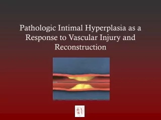
VASCULAR INJURY. Prof.Moaath AL Smady Jordan University Hospital. OUTLINE. DIAGNOSTIC MODALITIES COMPLICATIONS COMPARTMENT ANATOMIC EXPOSURE FEMORAL Vs POPLITIAL Vs SHANK Vs. PEREPHIRAL VASCULAE INJURY. Distal to Deltopectoral Groove Distal to Inginal ligament

E N D
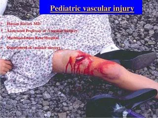
VASCULAR INJURY Prof.Moaath AL Smady Jordan University Hospital
OUTLINE DIAGNOSTIC MODALITIES COMPLICATIONS COMPARTMENT ANATOMIC EXPOSURE FEMORAL Vs POPLITIAL Vs SHANK Vs
PEREPHIRAL VASCULAE INJURY • Distal to Deltopectoral Groove • Distal to Inginal ligament • Hard signs of Vascular injury needs Surgery
Hard Signs • Observed pulsatile bleeding • Ongoing heamorrhage with shock • Arterial thrill by manual palpation • Bruit over or near the artery • Abscent distal pulse • Signs of distal ischemia • Visible expanding hematoma
Soft Signs • Significant hemorrhage by History • Small non expanding heamatoma • Decreased pulse compared to the contralateral extremity • Bony injury • Wound proximity (1 cm from Vs) • Neurologic abnormality(anatomicaly related nerve)
Doppler ultrasound • Ankle Brachial Index • ABI < 0.90 = 87% sensitive, 97% specific for arterial injury • In absence of hard signs, can substitute this for screeningarteriography
Doppler ultrasound • Determine presence/absence of arterial supply • Assess adequacy of flow PRESENCE OF SIGNAL DOES NOT EXCLUDE ARTERIAL INJURY !
Imaging StudyDuplex US • Reliable for • Injury to arteries and veins • A-V fistulas • Pseudoaneurysms • Thrombosis • *It has 95% sensitivity &99 % specificity
Imaging study CT Angio * Ct angiograph faster, less expensive and less invasive 90-100 % sensitivity and 98% - 100 %specificity * diagnostic study of choice *Limitations: • difficulty differentiating spasm from occlusion • artifact from high attenuation structures like bullet fragments or other foreign matter
Management • ABCs • Active bleeding, limb threatening ischemia OR • Stable, good limb viability may investigate • Non-operative management non-occlusive lesion in asymptomatic patient • Pre-operative management • Prophylactic antibiotic • Single dose heparin iv if no C/I • Do not reperfuse dead limb! amputation
Immediate treatment • Control bleeding • Replace volume loss • Cover wounds • Reduce fractures/dislocations • Splint • Re-evaluate
Option of vascular repair • Arterial repair: (1) direct arterial repair (2) arterial patch repair (3) interposition graft repair (4) bypass repair (5) ligation • Venous repair whenever possible avoid ligation.
Tension-free primary repair Primary repair defect < 1-2 cm
Venous injury • Should be repaired in stable patient if technically feasible • Lateral venorrhaphy, EEA • Complex repair (PTFE, SVG) • Patency 75% • Before arterial repair • Limb threatening ischemia Shunt a. repair v. • Ligation is safe alternative esp. in unstable patients, complex injuries. • 86% free of edema at D/C Yelon, et al. J Trauma 1992 • DVT 78%, no significant sequelae of CVI Kurtoglu, et al. Am Surg 2007 • Long term anticoagulation? • early arterial graft failure
What is the management ? Mangled extremity :injury that involve al least ¾ consisting of bone ,soft tissue ,vessel,nerves
Amputation • Non-viable or non-savagable limb • Irreversible limb ischemia • Safe life before limbs!!! • Amputation can be life saving in life threatening extremity bleeding • Functional outcome consideration
Hemorrhage • Thrombosis • Infection • Stenosis • Miscellaneous
THROMBOSIS • most important complication • relatively common compared with other complications, • Perry et al found an early occlusion rate of 9.1%,
Inadequate arterial de´bridement • A second adjacent injury • Residual distal arterial thrombus • Severe stenosis at the suture line • Undue tension due to significant missing arterial segment • Twisting,or too long graft to cause a kink or external compression of the graft
INFECTION • Primary skin closure in a war wound • Placement of a vascular graft in an area of established infection • Inadequate soft tissue de´bridement in an attempt to conserve tissue for coverage of a vascular repair • Inadequate de´bridement of a damaged vessel
STENOSIS • Technical complication • Tight suture repair. • Lateral repair without sufficient remaining wall • Residual arterial wall damage. • Tension on the suture line
MISCELLANEOUS COMPLICATIONS Acute • Errors in diagnosis second associated or adjacent arterial injury Improper identification of the arteries may occur • Edema • Embolization • Disseminated intravascular coagulopathies
Delayed • Chronic pain Drapanas et al (1970)7 found that chronic pain was a complaint in10.2% • Decreased function • Ischemic changes • Systemic complications • Arteriovenous fistulas and false aneurysms • Arteriosclerotic changes • Aneurysmal graft changes
Compartment Syndrome • occurs when muscle swells within osteofacial compartment pressure exceed capillary pressure they end up with ischemia • Causes • Distal pulses • Distal nerve • Examination • Pressure
Pain, aggravated during stretching of the muscle group involved. • Pressure. • Paresthesia. • Paralysis, late manifestation • Pulselessness very late stages • Pallor
Measurement of Pressures and Laboratory Tests • Intracompartmental Pressure Monitoring 40 • Serum CreatinePhosphokinase and Myoglobin • This enzyme indicates muscle necrosis. • not appropriate for early detection, • useful to monitor the progression of equivocal syndromes or recently decompressed compartments. • Pulse Oximetry and Near-Infrared Spectroscopy
TREATMENT • Adequate skin incision and an adequate fascial incision • Pharmacologic Interventions • Mannitol
FasciotomyFasciotomy to fully decompress all involved compartments is the definitive treatment for ACS in the great majority of cases
INDICATIONS FOR FASCIOTOMY • Prolonged hypotension • Swelling of the extremity • Extensive soft tissue damage • Combined venous and arterial injury • Combined bony plus arterial or venous injury or both • Delay between injury and definitive repair • Compartmental pressure 35 mm Hg
70% of all arterial • More than 90% penetrating • most resulting from GSWs.12 • Injuries to the femoral artery are not commonly associated with fractures of the femoral shaft
OPERATIVE MANAGEMENT • proximal injuries it is wise to initially expose the distal common iliac vessels through a separate incision control before entering the femoral triangle. • The length of the sterile field includes the entire abdomen to the toes in both lower • bleeding can be controlled by direct pressure from the source of bleeding • Blind clamping is strongly discouraged