Download
1 / 70
1.39k likes | 2.95k Vues
MRI Anatomy of the Shoulder. Functional Anatomy. Glenohumeral joint is a ball and socket synovial. Glenoid cavity inherently unstable. Stability provided by: Static constraints 3 glenohumeral ligaments glenoid labrum joint capsule Dynamic constraints, - rotator cuff muscles

E N D
Functional Anatomy • Glenohumeral joint is a ball and socket synovial. • Glenoid cavity inherently unstable. • Stability provided by: • Static constraints • 3 glenohumeral ligaments • glenoid labrum • joint capsule • Dynamic constraints, • - rotator cuff muscles • counteract the action of the deltoid by preventing the head of the humerus from moving superiorly when the arm is raised
The glenohumeral joint has the following supporting structures: • Superiorly • Coraco-acromial arch • Long head of the biceps tendon • Tendon of supraspinatus muscle • Anteriorly • Anterior labrum • 3 Glenohumeral ligaments • SGHL, MGHL, IGHL • (anterior band) • Subscapularis tendon • Posteriorly • Posterior labrum • Posterior band of the IGHL • Infraspinatus tendon • Teres minor tendon
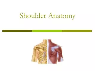
Articular surfaces The shoulder joint is composed of 3 bones and five articulations. Bones: Scapula Humerus Clavicle Articulations: Glenohumeral joint Acromio-clavicular joint Scapulothoracic joint Sternoclavicular joint Coracoclavicular joint
Joint capsule • Attached proximally to the glenoid labrum • Attached distally to the anatomical neck of the humerus • Capsule is thickened anteriorly by glenohumeral ligaments • Herniation of synovial membrane through an anterior defect in capsule & glenohumeral ligaments forms the subscapular bursa
Bursae Subdeltoid Between joint capsule and deltoid muscle Subcoracoid Between joint capsule and coracoid process Coracobrachial Between subscapularis and coracobrachialis Subacromial Between joint capsule and acromion Subscapular bursa Between joint capsule and subscapularis tendon
Rotator Interval • Triangular space between the supraspinatus and subscapularis tendons • Contains long head of biceps and SGHL • Acts to prevent anterior dislocation of the shoulder
Ligaments • 3 glenohumeral ligaments • Superior • Middle • Inferior • Coracohumeral ligament • Transverse humeral ligament • Between greater and lesser tuberosities of humerus, maintains long head of biceps in bicipital groove
Superior glenohumeral ligament Coracohumeral ligament Long head biceps tendon
Inferior glenohumeral ligament Axillary recess Subacromial-subdeltoid bursa
Glenoid labrum • The glenoid labrum is a fibrocartilaginous structure that attaches to the glenoid rim and is about 4 mm wide. • increases the superior-inferior diameter of the glenoid by 75% and the anterior-posterior diameter by 50% • Anteriorly, the glenoid labrum blends with the anterior band of the inferior glenohumeral ligament. • Superiorly, it blends with the biceps tendon and the superior glenohumeral ligament. • It is usually rounded or triangular on cross-sectional images.
Labral anatomy: Axial • The normal labrum demonstrates low signal intensity on all pulse sequences, due to the lack of mobile protons in this dense fibrocartilage. • On cross sectional imaging, the normal labrum is most commonly triangular, but can also be round, cleaved, notched, flat, or absent.
Labral Anatomy: Coronal • A fat suppressed oblique coronal T2-weighted MR image demonstrates homogeneously low signal intensity in the normal superior labrum.
Labral variants • These normal variants are all located in the 11-3 o'clock position.
It is important to recognise these variants, because they can mimick a SLAP tear. • These normal variants will usually not mimick a Bankart-lesion, since these are located at the 3-6 o'clock position, where these normal variants do not occur.
Sublabral recess Synovial recess between the superior labrum and the glenoid rim created by the attachment of the biceps tendon on the supraglenoid tubercle. Because of this recess, the labrum does not attach to the glenoid rim at the 12 o'clock position. There are 3 types of attachments of the superior labrum: Type I No recess between glenoid cartilage and labrum Type II Small recess. Type III Large sublabral recess.
Sublabral Foramen An unattached anterosuperior labrum at the 1-3 o'clock position. Anterior to biceps tendon It is seen in 11% of individuals. Not to be confused with a sublabral recess or SLAP-tear, which are also located in this region.
Differences between an sublabral recess and a SLAP-tear: A recess more than 3-5 mm is always abnormal and should be regarded as a SLAP-tear.
Buford complex Congenital labral variant 2 Features: Anterosuperior labrum is absent in the 1-3 o'clock position Middle glenohumeral ligament is usually thickened. It is present in approximately 1.5% of individuals.
On MR an os acromiale is best seen on superior axial images. Os Acromiale Results from failure of one of the acromial ossification centers to fuse. 5% of the population. Usually an incidental finding, regarded as a normal variant. May cause impingement because if it is unstable, it may be pulled inferiorly during abduction by the deltoid, which attaches here.
Acromion: 3 types Type 1 is a flat undersurface with a high angle of inclination. Type 2 is a curved arc and decreased angle of inclination. Type 3 is hooked anteriorly with a decreased angle of inclination.
The axillary artery begins at the lateral border of the first rib as a continuation of the subclavian artery. It changes its name to brachial artery at lower inferior border of the teres major muscle (8).
Axial • Deltoid • Anterior clavicular fibres – arise from superior anterior aspect of lateral clavicle • Lateral acromial fibres – arise from superior aspect of acromion process • Posterior fibres – arise from posterior border of spine of scapula
Axial Supraspinatus muscle Relatively small muscle Runs from the supraspinatous fossa of scapula to the greater tubercle of the humerus
Axis of supraspinatous tendon The supraspinatus tendon is the most important structure of the rotator cuff and subject to tendinopathy and tears. Tears of the supraspinatus tendon are best seen on coronal oblique and ABER-series. In many cases the axis of the supraspinatus tendon (arrowheads) is rotated more anteriorly compared to the axis of the muscle (yellow arrow). When you plan the coronal oblique series, it is best to focus on the axis of the supraspinatus tendon.
The attachments of the 3 rotator cuff muscles that insert onto the greater tubercle of the humerus can be abbreviated SIT when viewed from superior to inferior: Supraspinatus Infraspinatus Teres minor Sagittal post post SITS inlcudes Subscapularis which inserts onto the lesser tubercle of the humerus
Axial Pec major Deltoid anterior Pec minor Supraspinatus inserts most superiorly to greater tuberosity humeral head Subscapularis Infraspinatus Deltoid posterior
Infraspinatus (SIT) • Thick triangular muscle which occupies most of the infraspinatous fossa • Attaches medially to the infraspinatus fossa and laterally to the middle facet of the greater tubercle of the humerus • Trapezoidal insertion of infraspinatus onto humerus is much larger than the insertion of the supraspinatus
*Tip* Coronal oblique MRI • Supraspinatus – fibres run horizontally • Infraspinatus – fibres have a slightly oblique orientation
Subscapularis Large triangular muscle which fills the subscapular fossa Inserts onto the lesser tubercle of the humerus Subscapular fossa = anterior Infraspinatous fossa = posterior ant post
Axial Deltoid as one Coracobrachialis Coracobrachialis is the smallest of the three muscles that attach to the coracoid process. The other 2 muscles are pectoralis minor and biceps brachii. Distal insertion upper medial aspect of arm Teres minor
Teres minor(SIT) Origin: Superior part of lateral border of scapula Insertion: Inferior facet of greater tuberosity of humerus
Axial The short head of the biceps originates from the coracoid process (2) The long head originates from the supraglenoid tubercle (3)
Tendon of long head passes down along the intertubercular/bicipital groove of the humerus into the joint capsule Both heads arise on the scapula and join to form a single muscle belly which is attached to the upper forearm. Long head forms biceps-labral complex with superior glenohumeral ligament When the humerus is in motion, the tendon of the long head is held firmly in place in the bicipital groove by the greater and lesser tubercles and the overlying transverse humeral ligament.
Coronal Long head
Coronal Trapezius Posterior humeral circumflex artery and axillary nerve extends longitudinally from the occipital bone to the lower thoracic vertebrae and laterally to spine of the scapula Teres major Triceps
Teres major It arises from the dorsal surface of the inferior angle of the scapula Inserts onto intertubercular sulcus of humerus (Teres minor)
Coronal Infraspinatus Teres minor Triceps Teres major
Coronal Supraspinatus Subscapularis Teres major
Sagittal P A