Download
1 / 37
610 likes | 2.08k Vues
Synthesis and Characterization of Nanoparticulate Magnetic Materials. Villanova University, Villanova, PA 19085. Georgia C. Papaefthymiou. BuildMoNa Workshop University of Leipzig Leipzig, Germany October 28-29, 2010. Outline

E N D
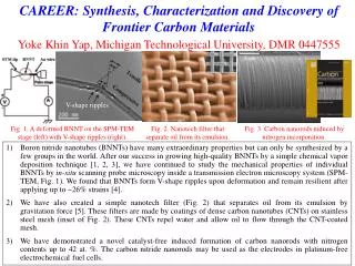
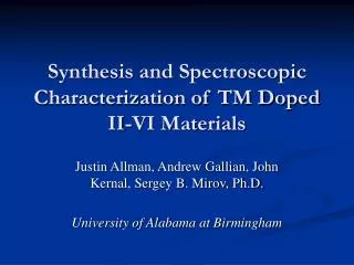
Synthesis and Characterization of Nanoparticulate Magnetic Materials Villanova University, Villanova, PA 19085 Georgia C. Papaefthymiou BuildMoNa Workshop University of Leipzig Leipzig, Germany October 28-29, 2010
Outline General concepts on the synthesis, stabilization and assembly of magnetic nanoparticles; examples Fundamentals of nanoparticle magnetism Macroscopic vs. microscopic magnetic characterization of Fe-based magnetic nanostructures; SQUID magnetometry, Mössbauer spectroscopy Isolated vs. interacting magnetic nanoparticles Conclusion Acknowledgements
Top → Down Synthesis by Physical Methods Up ← Bottom Synthesis by Chemical Methods Molecules Quantum behavior Nanoparticles Quantum-size effects Bulk Classical behavior Microscopic Mesoscopic Macroscopic 100 nm 1 nm Metal and Metal-Alloy Nanoparticles Metal and Metal-Oxide Nanoparticles • High Energy Ball Milling • Laser Ablation • Ion Sputtering • Thermal Evaporation • etc……. • Reduction of Metal Salts in Solution • Thermal Decomposition Reactions • Hydrolysis in Aqueous Solutions • Hydrolysis in Nonaqueous Solutions • etc……..
Nanoparticulate Magnetic Materials Nanostructured Nanocomposite matrix nanoparticle Abundance of grain boundaries Abundance of interfaces H. Gleiter, Acta Mater. 48, 1 (2000) Novel magnetic properties engineered through tailoring of the grain boundary or interfacial region and through interparticle magnetic interactions. Particles can interact via short-range magnetic exchange through grain boundaries or long-range dipolar magnetic interactions.
General Concepts in Nucleation and Growth of Magnetic Nanoparticles + Nucleation and Critical Radii DGn DGn = 4pr2DGs- (4/3)pr3DGv 0 r0 rc r Variation of Gibb’s free energy of nucleation with cluster radius during synthesis. rc is the kinetic critical radius and r0the thermodynamic critical radius Stabilization of nanoclusters of various size requires a competitive reaction chemistry between core cluster growth and cluster surface passivation by capping ligands that arrests further core growth. V. K. LaMer and R. H. Dinegar, J. Am. Chem. Soc. 72 (1950) 4847
Supramolecular Clusters Controlled hydrolytic polymerization of iron. Iron-core growth arrested via surface passivation with benzoate ligands. Observation of novel magnetic behavior. ~1 nm Fe11O6(OH)6(O2CPh)15.6THF Fe16MnO10(OH)10(O2CPh)20 G. C. Papaefthymiou, Phys. Rev. B 46 (1992) 10366 ~2 nm
Block Copolymer Nanotemplates Principles of synthesis Blocks of sequences of repeat units of one homopolymer chemically linked to blocks of another homopolymer sequence. Microphase separation due to block incompatibility or crystallization of one of the blocks. Templates for synthesis and arraying of metal oxide nanoclusters within space confined nanoreactors A-Block B-Block Chemical Link 0 - 21 % 21 - 34 % 34 - 38 % 38 - 50 % Increasing Volume Fraction of Minority Component
Cobalt Ferrite Nanocluster Formation within Block Copolymers G.C. Papaefthymiou, S.R. Ahmed and P. Kofinas Rev. Adv. Mater. Sci. 10 (2005) 306
Transmission Electron Microscopy CoFe2O4 Block Copolymer Films Morphology of block copolymer films: ensemble of polydispersed CoFe2O4 nanoparticles, oval in shape and of average diameter of 9.6 ± 2.8 nm. Ahmed, Ogal, Papaefthymiou, Ramesh and Kofinas, Appl. Phys. Letts 80 (2002) 1616
Self-assembly within Protein Cages: Ferritin Apoferritin Ferritin 24 amino acid subunits form a robust protein cage → ←7nm → The Ferroxidase and Nucleation sites of Human H-chain Ferritin → → Iron Mineralization in Ferritin ferrihydrite 2Fe2+ + O2 + 4H2O →2FeOOH (core) + H2O2 + 4H+ (1) 4Fe2+ + O2 + 6H2O →4FeOOH (core) + 8H+ (2) 2Fe2+ + H2O2 + 2H2O →2FeOOH (core) + 4H+ (3) Demineralization followed by metathesis mineralization leads to biomimetic synthesis of various nanoscale particles. A large number of nanostructures and mono-layer films on various supports have been produced including metal oxide (Fe3O4, Co3O4), iron sulfide, metallic (Co, Mn, U, Co/Pt, Ni, Cr, Ag) and semi-conducting (CdS, CdSe) structures, and FeOOH•(MO4)x, where M=P, As, Mo or V.
Two-dimensional Array of Ferritin Ensemble of monodispersed magnetic nanoparticles I. Yamashita Thin Solid Films, 391 (2001) 12
Monodispersed γ-Fe2O3 nanoparticles Thermal decomposition of iron pentacarbonyl, Fe(CO)5, in the presence of oleic acid produced monodispersed metal iron particles. Controlled oxidation using trimethylamine oxide, (CH3)3NO, as a mild oxidant produced highly crystalline γ-Fe2O3 particles. The particles were in the size range 4 nm to 16 nm diameter depending on experimental conditions. Highly uniform, oleic acid covered, magnetic nanoparticles of γ-Fe2O3, ~(11.8 ± 1.3) nm diameter are shown. XRD patterns confirm the presence of Fe2O3. D.K. Yi, S.S. Lee, G.C. Papaefthymiou, J.Y. Ying, Chem. Mater. 18 (2006) 614
Schematic of the synthesis of MP/SiO2/MS nanoarchitectures MP = Magnetic Particle SiO2 =Solid Silica MS = Mesoporous Silica D.K. Yi, S.S. Lee, G.C. Papaefthymiou, J.Y. Ying, Chem. Mater. 18 (2006) 614
Solid-silicacoatedγ-Fe2O3 nanoparticles TEM micrographs of ~12 nm γ-Fe2O3 particles covered with solid silica shell. Shell thickness from 1.8 nm to 25 nm was achieved. Scale bar 20 nm D.K. Yi, S.S. Lee, G.C. Papaefthymiou, J.Y. Ying, Chem. Mater. 18 (2006) 614
Higher Nanoarchitectures TEM micrographs of γ-Fe2O3 core-solid silica shell-mesoporous silica shell nanocomposites • ~ 12 nm maghemite particles were used as templates • A thick mesoporous layer (~21nm) was obtained using a mixture of TEOS and C18TMS, 260 μl and • a thinner mesoporous layer (~10nm) was obtained using a mixture of TEOS and C18TMS, 120 μl. • In both cases, (a) and (b), ca. 25 nm solid silica shell coated Fe2O3 core-solid silica shell nanocomposites were used as templating cores.
Fundamentals of Magnetic Ordering Indirect Exchange Direct Exchange Magnetic ordering in solids is due to Quantum Mechanical Exchange and the Pauli Exclusion Principle Bethe-Slater Curve Curie temp Tcin °C, Iron (Fe) 770, Cobalt (Co) 1130, Nickel (Ni) 358, Iron Oxide (Fe2O3) 622
Magnetic Anisotropy Minimization of magnetostatic energy Bulk Co in its demagnetized, multi-domain state leads to domain wall formation ↘ Uniaxial Magnetic Anisotropy Anisotropy Field Moment rotation at a Bloch Wall Exchange energy per unit area of Bloch wall where for a simple cubic lattice with lattice constant a.
Hard process in a single domain system Process of Magnetic Saturation of a Multi-domain Particle Easy process in a multi-domain system Hysteresis Loop The hysteresis loop defines the technological properties of the magnetic material Saturation Magnetization Remnant Magnetization Coercivity
Critical Size for SMD Particles Magnetostatic vs. wall energy as a function of particle size for a spherical particle of radius r ← Below Rc the particle is a Single Magnetic Domain, and thus permanently magnetized. The demagnetized state cannot be formed. ← Rc~ 100 nm
Coercivity as a function of particle size F. E. Luborsky J. Appl. Phys.32 (1961) S171
Nanomagnetism: Coercivities and Spin Reversal Mechanisms S-D M-D SP Maximum coercivity Unstable Hc Dp 0 Ds Particle Diameter D Multi-magnetic domain structure Magnetic wall movement Single-magnetic domain particle Coherent spin rotation Nanoparticle K ~ 105 J/m3 Bulk K ~103 J/m3
Origin of magnetic anisotropy enhancement in nanoparticles F. Bødker, S. Mørup, S. Lideroth, Phys. Rev. Lett. 72 (1994) 282 c = core s = surface σ = stress sh = shape
Nanoparticle coercivity for coherent spin rotation (Stoner and Wohlfarth model) Maximum coercivity for coherent spin rotation of a single magnetic domain particle with uniaxial total effective anisotropy coherent moment rotation E.C. Stoner, E.P. Wohlfarth, Trans. Roy. Soc. Lond. A 240 (1948) 599
Spin Dynamics in Magnetic Nanoparticles Easy axis Temperature dependence of coercivity Superparamagnetic relaxation time (thermally assisted spin reversals) Due to fast moment reversals at elevated temperatures the internal magnetic order of the particle escapes detection. You must either lower the temperature or use ultrafast measuring techniques that can record the moment before it flips.
Superparamagnetism of Small Magnetic Particles Energy barrier Δ E = KuV where Ku is the effective uniaxial magnetic anisotropy Energy density and V is the particle volume Relaxation Time tRELAX = t0 exp (KuV/kΤ) Magnetocrystalline Anisotropy Observe net magnetic moment when tMEAS < tRELAX Shape Anisotropy Surface effects
Micro-magnetics and Spin Dynamics -Mössbauer spectroscopic measurements Probe local magnetic moments and internal magnetic fields, with a response time of tm = tMöss = 10 ns -DC Magnetization measurements Probe global magnetic properties in an applied field,with a response time of τm = τSQUID = 10 s
Hysteresis Loops for CoFe2O4 Block Copolymers Hysteresis due to particle moment rotation away from the particle’s easy axis to the direction of the applied magnetic field. The temperature at which the coercivity vanishes defines the blocking temperature TBfor SQUID magnetometry. Ahmed, Ogal, Papaefthymiou, Ramesh and Kofinas, Appl. Phys. Letts 80 (2002) 1616
Nuclear Hyperfine Interactions with Mössbauer Spectroscopy Observed Spectrum Observed Effect Illustration Isomer Shift Interaction of the nuclear charge distribution with the electron cloud surrounding the nuclei in both the absorber and source. v 0 Quadrupole Splitting Interaction of the nuclear electric quadrupole moment with the EFG and the nucleus v 0 Zeeman Effect (Dipole Interaction) Interaction of the nuclear magnetic dipole moment with the internal magnetic field on the nucleus. I(v) v 0
Modeling Dynamical Spin Fluctuations in Isolated Nanostructures Mössbauer spectra of lyophilized, in vitro reconstituted HoSF ferritin. 80 K Determination of Blocking Temperature Experimentally the temperature at which the Mössbauer spectra pass from magnetic, six-line spectra to paramagnetic or quadrupolar, two-line spectra defines TBfor Mössbauer 40 K TB = 40 K Theoretically TBis defined by: 30 K Absorption (arb. units) → 25 K Spectrum Key Magenta: spectral signature of magnetic particle core (internal iron sites) Green: spectral signature of surface layers (surface iron sites) 4.2 K Velocity (mm/s) G. C. Papaefthymiou, Biochim. Biophys. Acta 1800 (2010) 886 G. C. Papaefthymiou, et. al.MRS Symp. Proc. Fall 2007
Zero-field cooled and field-cooled magnetization of lyophilized HoSF ferritin 25-nm thick protein shell FC ZFC Typical ZFC/FC behavior of an ensemble of magnetically isolated superparamagnetic particles Note: Saturation magnetization is ~ 0.05 emu/g, weakly magnetic.
Determination of Kufor an ensemble of superparamagnetic nanoparticles • Determine average particle volume <V> by TEM • Determine TBwith two different techniques, whose measuring response times lie in different time windows • Use the Arrhenius equation above to determine τ0and Ku
Surface Effects:Temperature Dependence of Mössbauer Magnetic Hyperfine Fields 80 K CME model, double potential well 40 K 30 K 25 K 4.2 K complex potential energy landscape at the surface Velocity (mm/s) Collective magnetic excitations below TB S. Mørup and H. Topsøe, Appl. Phys. 11 (1976) 63
Mössbauer Spectra of γ-Fe2O3/Solid Silica Nanoarchitectures Bare 12 nm particles 12 nm particles with 25 nm SiO2 shell Spectral Key: Blue A-sites, Green B-sites of spinel structure G.C. Papaefthymiou et. al. Phys. Rev. B 80 (2009) 024406
Effect of silica shell on the RT Mössbauer Spectra Behavior typical of strongly interacting particles Bare γ-Fe2O3 nanoparticles γ-Fe2O3 nanoparticles with 4 nm silica shell γ-Fe2O3 nanoparticles with 25 nm silica shell G.C. Papaefthymiou et. al. Phys. Rev. B 80 (2009) 024406
Magnetization of γ-Fe2O3/Solid Silica/Mesoporous Silica Nanoarchitectures A-bare * B-4 nm (S) C-25 nm (S) D-25 nm (S) + 10 nm (MS) E-25 nm (S) +21 nm (MS) Typical behavior of strongly interacting magnetic nanoparticles, spin-glass-like systems. * Bare particles are covered with a very thin layer (~1 nm) of oleic acid. Saturation magnetization of the order of ~ 8 emu/g, strongly magnetic
Conclusion Ferrihydrite is an antiferro-magnet. Magnetization of ferritin is due to uncompensated spins at the surface → Weak magnetism. Protein coat of only 2.5 nm thickness sufficient to magnetically isolate the ferritin iron cores Maghemite is a ferri -magnet due to uncompensated spin sublattices in its spinel structure. In small particles uncompensated spins at the surface also contribute → Strong magnetism. Silica coat of 23 nm thickness insufficient to isolate the γ-Fe2O3 cores Dipole-dipole interaction ~
Acknowledgements Steve Lippard, MIT Peter Kofinas, University of Maryland Dennis Chasteen, University of New Hampshire Jackie Ying, IBN Singapore Eamonn Devlin, NCSR Demokritos, Greece NSF, EU/Marie-Curie