Download
1 / 82
950 likes | 1.17k Vues
NEONATAL INTESTINAL OBSTRUCTION. DR ENIVWENAE OGHENEKARO. OUTLINE. INTRODUCTION EMBYOLOGY CAUSES OF NEONATAL INTESTINAL OBSTRUCTION PATHOGENESIS CLINICAL MANIFESTATION INVESTIGATIONS TREATMENT FOLLOW – UP CONCLUSION. INTRODUCTION.

E N D
NEONATAL INTESTINAL OBSTRUCTION DR ENIVWENAE OGHENEKARO
OUTLINE • INTRODUCTION • EMBYOLOGY • CAUSES OF NEONATAL INTESTINAL OBSTRUCTION • PATHOGENESIS • CLINICAL MANIFESTATION • INVESTIGATIONS • TREATMENT • FOLLOW – UP • CONCLUSION
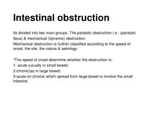
INTRODUCTION • A neonate is a baby (newborn) within the first 28 days of life. • Intestinal obstruction is stoppage of the onward (craniocaudal) passage of intestinal contents. • Neonatal intestinal obstruction is the commonest neonatal surgical emergency. • It can be life threatening and requires prompt recognition and intervention.
INTRODUCTION • Delicate physiological adjustment to extra-uterine life. • High risk for surgery and anaesthsia • Improvements in surgery, anaesthesia, neonatal intensive care facilities and parenteral nutrition has improved outcome • Still a challenge in the developing countries
INTRODUCTION • Delicate physiological adjustment to extra-uterine life. • Congenital anomaly may be associated with intrauterine growth restriction and preterm delivery. • Adaptive mechanisms further impaired in preterm, LBW, and SGA neonates
INTRODUCTION • Problems in the neonate compounded by surgical pathology • Hypothermia • Hypoglycaemia • Susceptibility to infection • Jaundice • Respiratory embarrassment • Vitamin K deficiency bleeding
Relevant Embryology • 3rd week of development: formation of the gut tube from the yolk sac
Relevant Embryology • End of 4th week of development: formation of the gut tube from the yolk sac
Relevant Embryology • 5th week of development: formation of the gut tube from the yolk sac
Relevant Embryology • 6th week of development: formation of the gut
Relevant Embryology • Duodenum: solid stage (5th/6th week), Recanalization (11th week)
Relevant Embryology • Development of the pancreas (4th/5th week)
Relevant Embryology • Development of the pancreas (4th/5th week)
Relevant Embryology • 6th – 10th week of gut development: Physiologic herniation
Relevant Embryology • 10th – 12th week of gut development: Return of the midgut
Relevant Embryology • Hindgut development
Relevant Embryology • Innervation: Migration of neural crest cells; 5th – 12th week
Relevant Embryology • Blood supply
Aetiology of Neonatal Intestinal obstruction • Acquired • Congenital
Aetiology of Neonatal Intestinal obstruction • Acquired • Medical • Dyselectrolytaemia • Hypothyroidism • Hypothermia • Sepsis • Necrotizing enterocolitis • Surgical • Complicated Necrotizing enterocolitis • Intussusception
Aetiology of Neonatal Intestinal obstruction – Congenital • DUODENAL • Atresia • Stenosis • Annular pancreas • Malrotation • JEJUNOILEAL • Atresia • Meconium Ileus • Malrotation • Vitelineanormalies • Congenital bands • Obstructed inguinal hernias • COLORECTAL • Hirschsprung’s disease • Anorectal malformation • Meconium plug • OTHERS • Intra-abdominal masses , abdominal wall defects, intestinal duplications, small left colon syndrome
Aetiology of Neonatal Intestinal obstruction – Congenital • Common Causes: • Anorectal malformation • Hirschsprung’s disease • Duodenal atresia • Jejunoileal atresia • Malrotation with midgut volvulus • Obstructed inguinal hernia
PATHOPHYSIOLOGY OBSTRUCTION PROXIMAL ACCUM DISTENSION INCREASE PERISTALSIS NO INTERVENTION WANES (ILEUS)
PATHOPHYSIOLOGY STASIS BACTERIAL PROLIFERATION TRANSLOCATION / TRANSMIGRATION CIRCULATION PERITONITIS SEPTICEMIA
PATHOPHYSIOLOGY DISTENSION VILLOUS ATROPHY ABSORPTION SECRETION VOMITING E/U IMBALANCE
PATHOPHYSIOLOGY INTRALUMINAL PRESSURE ABOVE VASCULAR PRESSURE ISCHAEMIA NECROSIS PERFORATION GENERALIZED PERITONITIS
PATHOPHYSIOLOGY • Constipation • Bowel distal to obstruction - peristalsis eventually stops after expelling contents
PATHOPHYSIOLOGY • Duodenal Atresia • Incidence: 1:5000 – 10000 • Male > Female • Failure of recanalization during the embryonic period • Classification: Types I, II & III
PATHOPHYSIOLOGY • Duodenal Atresia • 85% postampullary, 15% preampullary • Vomiting (bilious) form 1st day of life • Maternal polyhydramnios • More than 50% associated anomalies • Down’s syndrome, cardiac defects, GIT, prematurity, etc
PATHOPHYSIOLOGY • Jejunoileal Atresia • Incidence of 1: 5000 • M:F = 1:1 • 10% Associated anomalies • Likely due to a vascular disruption, leading to resorption of affected segment of ischaemic bowel.
PATHOPHYSIOLOGY • Jejunoileal Atresia • Classified as Type I, II, IIIa, IIIb and IV. • Polyhydramnios with proximal bowel segment • May pass meconium
PATHOPHYSIOLOGY • Malrotation Syndrome • Related to problems associated with midgut rotation and fixation • Incidence of 1:6000 • Up to 75% present in the neonatal period
PATHOPHYSIOLOGY • Malrotation Syndrome • Spectrum • Non rotation • Incomplete Rotation • Hyper-rotation • Reversed rotation • Encapsulated small intestines • Internal Hernias (Paraduodenal & Mesocolic) • Ladd’s bands (Extrinsic duodenal obstruction)
PATHOPHYSIOLOGY • Malrotation Syndrome: midgutvolvolus most feared complication
PATHOPHYSIOLOGY • Anorectal Malformation • Absent normal anal opening • Abnormal development of urorectal septum resulting in incomplete separation of cloaca into urogenital & anorectal portions. • Incidence of 2 -2 .5:10,0000 • M:F = 1 – 1.8:1 • Associated anomalies • VACTERL-H
PATHOPHYSIOLOGY • Anorectal Malformation • Associations • V = Vertebral • A = Anorectal • C = Cardiac • T = Tracheo-oesophageal fistula • E = Esophageal • R = Renal • L = Limb • H = Hydrocephalus
ANORECTAL MALFORMATION – LOW ARM WITH BUCKET HANDLE DEFORMITY
PATHOPHYSIOLOGY • Hirschsprung’s disease • Functional intestinal obstruction caused by the congenital absence of parasympathetic ganglion cells of the ENS in the distal colon. • 1 in 5000 live births • M:F = 4:1 • Asians > Whites > Blacks • Sporadic : Hereditary = 90 : 10
HD Classification • Ultrashort segment HD -----------------<1% • Short segment HD ------------------------70-80% • Long segment HD -------------------------10-25% • Total colonic aganglionosis (TCA) -----3-15% • Total intestinal aganglionosis (TIA)---0.4-4
PATHOPHYSIOLOGY • Hirschsprung’s disease • May present as: • Acute neonatal intestinal Obstruction • Chronic large bowel obstruction • Acute on chronic large bowel obstruction • Hirschsprung’s Associated Enterocolitis (HAEC) • History of delayed passage of meconium • Constipation from birth
PATHOPHYSIOLOGY • Obstructed Inguinal Hernias • Inguinal hernia is present in upto 10 – 30% of preterm and 3- 5% of term babies • Likelihood of obstruction and strangulation is higher in this group compared to older children and adults. • With delayed intervention, bowel loops may herniate into the patent processusvaginalis, get incarcerated in the inguinal canal, may become obstructed and may get strangulated
PATHOPHYSIOLOGY • Obstructed Inguinal Hernia
Clinical Presentation - History • Abdominal pain – inconsolable cry • Refusal of feeds • Vomiting • Failure to pass meconium • Constipation • Abdominal Swelling • Passage of blood in stool • Groin swelling • Absent/ abnormal anal opening • Perineal fistula, or passage of meconium/ flatus in urine?
Clinical Presentation - History • Convulsion • Fever • Difficulty with breathing • Reduced volume/frequency of micturition • Jaundice
Clinical Presentation - History • Family history • Maternal History • Assisted reproduction • Drugs – teratogens, vasoactive drugs, herbal medications • History suggesting polyhydramnios • Routine Ultrasound scan • Delivery • Resuscitation • Other associated anomalies • Treatment so far