Download
1 / 1
10 likes | 104 Vues
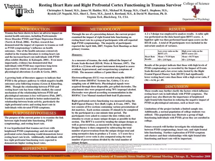
Resting Heart Rate and Right Prefrontal Cortex Functioning in Trauma Survivors. Christopher S. Immel, M.S., James M. Hadder, M.S., Michael M. Knepp, M.S., Chad L. Stephens, M.S., Ryoichi J.P. Noguchi, M.S., Sheri L. Towe, M.S., Jared A. Rowland, M.S., & David W. Harrison, Ph. D.

E N D
Resting Heart Rate and Right Prefrontal Cortex Functioningin Trauma Survivors Christopher S. Immel, M.S., James M. Hadder, M.S., Michael M. Knepp, M.S., Chad L. Stephens, M.S., Ryoichi J.P. Noguchi, M.S., Sheri L. Towe, M.S., Jared A. Rowland, M.S., & David W. Harrison, Ph. D. Virginia Tech, Blacksburg, VA, USA. INTRODUCTION METHODOLOGY RESULTS Trauma has been shown to have an adverse impact on mental health outcomes, including Posttraumatic Stress Disorder PTSD) and Major Depression Disorder (Norris & Elrod, 2006). Further, research has demonstrated the impact of exposure to trauma as well as PTSD symptomology’s influence on health outcomes. One of the health outcomes extensively studied in the trauma literature is resting heart rate and the increased heart rate individuals with PTSD often exhibit (Buckley & Kaloupek, 2001). Even more importantly, evidence has demonstrated that individuals with PTSD may experience long-term hyperactivity which can result in permanent physiological alterations (Lovallo & Gerin, 2003). A growing body of literature appears to indicate that PTSD is the primary pathway to which negative health outcomes arise following trauma (Green & Kimerling, 2004). Though the relationship between PTSD and resting heart rate has been widely studied, the causal mechanism of the relationship between PTSD and physiological outcomes is less well known (Pole, 2007). Further research is needed to better understand the relationship between brain activity, particularly the right prefrontal cortex and resting heart rate in individuals with elevated PTSD symptoms. A 2 x 3 design was employed to analyze results. A cubic split was performed on the data based upon RFFT scores. A high/low split was then performed based upon scores on the IES-R scale. A total of 38 participants were included in the univariate analysis of variance. Results of the project indicate that those with high levels of PTSD symptomology (as recorded by the Impact of Events Scale-Revised) and a low error ratio (tested by the Right Frontal Figural Fluency Task [RUFF]) had significantly lower resting heart rates than those with a high error ratio, F (36) = 6.34, p< .05. Through the use of a preexisting dataset, the current project examined the impact of right frontal lobe functioning on resting heart rate in a group of participants with self-reported high PTSD symptomology. The majority of participants reported the April 16th, 2007, Virginia Tech Shootings as their primary trauma. Measures: As a measure of trauma, the study utilized the Impact of Events Scale-Revised (IES-R; Weiss & Marmar, 1997). The IES-R is a 22 item self-report instrument designed to assess avoidant, hyperarousal, and reexperiencing symptoms of PTSD. The measure utilizes a 5 point likert scale. Electrocardiogram (ECG) was recorded using the BIOPAC MP30 system (BIOPAC Systems Inc, Goleta, CA) at a sampling rate of 1000 hertz. Physiological signals were acquired through three disposable, pre-gelled electrodes. The attachment sites were prepared using 70% isopropyl alcohol. Electrodes were placed according recommendation in the BIOPAC User Manual standard limb lead II placement. Right prefrontal cortex functioning was measured using the Ruff Figural Fluency Test (Ruff, Light, & Evans, 1987). This test consists of five sections containing various presentations of dot matrices. Each section contains 35 stimuli to be completed by the participant in a one-minute time period. The participants were asked to connect the dots within each stimulus to create as many unique designs as possible in that section. The RFFT is scored by totaling the unique designs as well as perseverations, as measured by design repetitions. Design fluency is then computed by subtracting the total number of perseverations from the unique design total and using normative data to produce a T score. A T score for a participant’s error ratio is also computed using the total number of perseverations divided by the total number of unique designs. DISCUSSION These results may further clarify the factors which influence resting heart rate in individuals with PTSD symptoms. The project may also illustrate the importance of heightened right frontal lobe function in reducing the negative impact of PTSD on physiological outcomes, such as heart rate. Limitations of the project include a limited sample size. Additionally, a normal population of college students was utilized. This population may illustrate a group of high functioning individuals with PTSD, given they are enrolled in college. Future research will further investigate the relationship between PTSD symptomology, heart rate, and right frontal lobe functioning. Further exploration of PTSD symptom sub-clusters and their relationships with right frontal lobe functioning and heart rate will be important. PURPOSE/HYPOTHESES The purpose of the current poster is to examine the link between right frontal lobe functioning, PTSD symptomology, and resting heart rate. It was hypothesized that trauma survivors with heightened PTSD symptomology and elevated right prefrontal cortex functioning would demonstrate lower resting heart rate levels. Additionally, individuals with less right prefrontal functioning were expected to demonstrate higher resting heart rates. REFERENCES -Contact Information: Christopher Immel via immel@vt.edu. International Society of Traumatic Stress Studies 24rd Annual Meeting, Chicago, IL, November 2008