Download
1 / 16
160 likes | 463 Vues
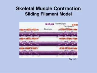
Sliding Filament Mechanism. Ch. 10-3 notes. Sliding Filament Mechanism. Describes the cause and mechanism of muscle contraction Myosin heads attach to and “walk” along the thin filaments at both ends of a sarcomere

E N D
Sliding Filament Mechanism Ch. 10-3 notes
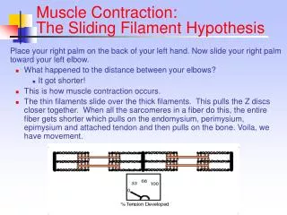
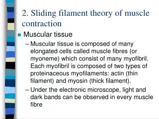
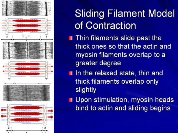
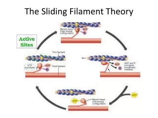
Sliding Filament Mechanism • Describes the cause and mechanism of muscle contraction • Myosin heads attach to and “walk” along the thin filaments at both ends of a sarcomere • The thin filaments are pulled toward the M line and meet at the center of the sarcomere • The Z discs come closer together and the sarcomere shortens. • This shortening shortens the entire muscle
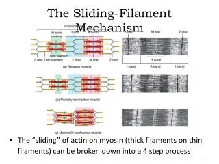
The Contraction Cycle • Contraction • SR releases Ca2+ ions into the cytoplasm • Ca2+ binds to troponin to move tropomyosin out of the way of the myosin-binding sites on actin • Contraction cycle – repeating sequence of events that causes the filaments to slide • 4 steps
4 Steps to Contraction Cycle 1. ATP hydrolysis • Breaking ATP into ADP + P adds energy to the myosin head so it can turn 2. Attachment of myosin to actin to form cross-bridges • Myosin head attaches to binding site on actin • Releases P • Crossbridge forms
4 Steps to Contraction Cycle 3. Power Stroke • Myosin crossbridges rotate 4. Detachment of myosin from actin • Myosin remains attached until another ATP binds causes it to detach • * rigor mortis Contraction cycle continues - with ATP - with Ca2+ levels high enough
Contraction • The myosin heads “walk” down the actin • They can attach and detach about 5 times per second http://www.dnatube.com/video/4154/Muscle-Contraction http://www.dnatube.com/video/1305/Muscle-Contraction http://www.dnatube.com/video/1306/Muscle-contraction http://www.dnatube.com/video/1308/Muscle-Contraction http://www.sci.sdsu.edu/movies/actin_myosin_gif.html
Neuromuscular Junction (NMJ) • Somatic motor neurons – neurons that stimulate skeletal muscle fibers • Place where action potentials arise • Synapse – region where 2 neurons or 1 neuron and a target cell (muscle fiber) meet • Synaptic cleft – gap that separates two cells • Neurotransmitter – chemical released for the 2 cells to communicate
NMJ Parts • At the end of each axon are synaptic vesicles filled with acetylcholine (ACh) • ACh is the neurotransmitter • Motor end plate – sarcolemma opposite the synapse • Contains acetylcholine receptors • Action potential starts here with stimulation from neurons
Action Potential 1. Release of ACh • Vesicles do exocytosis • Diffuse to motor end plate 2. Activation of ACh receptors • 2 Ach bind to motor end plate and opens the receptor • Na+ flows across membrane through channel
Action Potential 3. Production of muscle action potential • Na+ makes fiber more + charged • This causes release of Ca+ • Contraction cycle! 4. Termination of ACh activity • Acetylcholinesterase (AChE) – breaks down ACh so further muscle contractions don’t occur • Unless more ACh is released