Download
1 / 21
330 likes | 472 Vues
The adrenal cortex. Clinical biochemistry 200 8 (J.S.). C 27. Cytochrome P450 monooxygenases control the steroidogenesis:

E N D
The adrenal cortex Clinical biochemistry 2008 (J.S.)
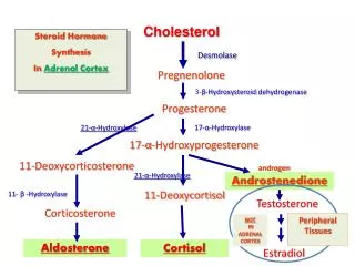
C27 Cytochrome P450 monooxygenasescontrol the steroidogenesis: The rate limiting step of the biosynthetic pathway that generates C21steroids is catalysed by P450scc(side-chain cleaving cholesterol 20,22-desmolase). (The dehydrogenation / isomerization is catalysed by 3β-hydroxysteroid dehydrogenase.) Three sequential hydroxylations at C-21, C-17α, and C-11β are catalysed by P450c21, P450c17, P450c11. Unique is the hydroxylation / dehydrogenation at C-18catalysed by P450 aldosterone synthase. The splitting at C-17 that generates C19 steroids is catalysed by P450c17 17-20 lyase / desmolase. Cholesterol (cholest-5-ene-3β-ol)is the precursor for all steroid hormone synthesis.
Steroidogenesis CHOLESTEROL Pregnenolone17-OH pregnenoloneDHEA (dehydroepiandrosterone) Progesterone 17-OH progesteroneANDROSTENDIONE DEOXYCORTICOSTERONE11-Deoxycortisol CorticosteroneCORTISOL 18-OH corticosterone ALDOSTERONE Gonades and peripheral tissues TESTOSTERONE Adrenal cortex - zona glomerulosa Cortisone ESTRADIOL
Enzymes catalysing the corticosteroid synthesis: Cholesterol 17-Hydroxylase / 17,20-lyase Chol. side chain cleavage enzyme Pregnenolone 17-Hydroxypregnenolone DHEA 3-Hydroxysteroid dehydrogenase PROGESTERONE 17-Hydroxyprogesterone Androstenedione 21-Hydroxylase DOC 11-Deoxycortisol 11-Hydroxylase Corticosterone CORTISOL Aldosterone synthase 18-Hydroxycorticosterone Synthesis is enabled by small numberof multifunctional enzymes. Aldosterone synthase ALDOSTERONE
Glucocorticoids Adrenal androgens Mineralocorticoids Catecholamines Medulla In adrenal cortex, there are three distinct cellular zones differing in the enzymatic equipment. The three zones form two functional units that are controlled independently. The cells of the outer layer – zona glomerulosa – do not express 17α-hydroxylase so that they do not produce the precursors of glucocorticoids and adrenal androgens. On the other hand, the gene for aldosterone synthase is expressed only in that zone – the unique site of aldosterone production. The synthesis and secretion of aldosterone is controlled by renin-angiotensin systém; the influence of adrenocorticotropin (ACTH) is very weak and transient. Both inner zones of adrenal cortex – zona fasciculata and reticularis – produce glucocorticoids and androgens, the production of less effective mineralocorticoids deoxycorticosterone (DOC) and corticosterone is not very important. The synthesis and secretion is controlled by ACTH.
Synthesis of aldosterone in zona glomerulosa of adrenal cortex CHOLESTEROL Pregnenolone Progesterone DOC Corticosterone 18-OH corticosterone ALDOSTERONE Chol. side chain cleavage enzyme ANGIOTENSIN II Carboxypeptidase ACE 3-Hydroxysteroid dehydrogenase ANGIOTENSIN I 21-Hydroxylase RENIN (proteinase) 11-Hydroxylase ANGIOTENSINOGEN (plasma 2-globulin) Aldosterone synthase Secretion of the juxtaglomerular cells is stimulated by - pressure decrease in arteriolae afferentes - circulating catecholamines - the decrease of [Na+] + [Cl-] in the tubular liquid (detected by cells of macula densa) Aldosterone synthase REABSORPTION of Na+, excretion of K+
Aldosterone 11,18-epoxy-18,21-dihydroxypregn-4-ene-3,20-dione
Synthesis of steroids in zona fasciculata and zona reticularis: CHOLESTEROL Pregnenolone 17-OH pregnenoloneDHEA Progesterone17-OH progesteroneANDROSTENDIONE DOC11-Deoxycortisol CorticosteroneCORTISOL Chol. side chain cleavage enzyme 17-Hydroxylase / 17,20-lyase 3-Hydroxysteroid dehydrogenase 21-Hydroxylase 11-Hydroxylase NEGATIVE FEEDBACK CONTROL CORTICOLIBERIN (CRF) ACTH (CORTICOTROFIN)
Cortisol (hydrocortisone) 11,17,21-trihydroxypregn-4-ene-3,20-dione
Assessment of adrenocortical function Basal investigation - glucocorticoids Serum cortisol (total) – basal value at 08:oo AM men 250 – 650 nmol/l, women 140 – 740 nmol/l. Cortisol - circadian rhythm – disturbed if at 05:oo PM more than 410 nmol/l. Free urinary cortisol - range 14 – 135 nmol/d (HPLC) 50 – 250 nmol/d (RIA) - mineralocorticoids Serum Na+, K+, daily output U- Na+, K+, FE (Na+,K+). Plasma renin activity, PRA – the method is not quite specific, though proportional to plasma angiotensin I concentrations; in blood samples taken after entire bed-rest, the activity is less than 2 nmol/h per litre – positional changes or stress readily increase the PRA.. Captopril test is very useful in the diagnosis of renovascular hypertension: On the contradistinction to essential hypertension, a distinct increase in PRA can be observed 1 hour after application of captopril (an inhibitor of angiotensin converting enzyme, ACE). Screening for 17α-hydroxyprogesterone in newborns from dry blood drop to detect dangerous salt-wasting form (a serious hypoaldosteronism) of 21-hydroxylase deficit..
Dynamic function tests Short ACTH stimulation test by i.m. injection of 250 µg synacthen (synthetic ACTH): Serum cortisol is measured before and at 30 and 60 min after injection. Adrenocortical insufficiency is excluded, if an increase over 690 nmol/l is observed.. Insulin stimulation test (insulin tolerance test). Hypoglycaemia (2.6 mmol/l or 50 % of initial value) causes release of CRH, ACTH, and cortisol. If an increase of serum cortisol is observed during 2 hours after i.v. injection of 0.1 IU/kg, adrenocortical insufficiency or decrease in pituitary ACTH reserve are excluded. Dexamethasone suppression tests. One-day screening test: Blood sample for cortisol determination in the morning, 1 mg dexamethasone perorally at bedtime, blood sample for cortisol the following morning at 08:oo AM. Normally, serum cortisol being less than 100 nmol/l, insufficient suppression in Cushing's syndrome. Two-day high-dose test: Basal serum cortisol and urinary cortisol excretion. Dexamethasone 2 mg is taken 6-hourly for 2 days, serum and excreted cortisol is measured. In healthy individuals, serum cortisol < 55 nmol/l, excreted < 55 nmol/d. Metyrapone blocking test(inhibition of 11-hydroxylase causes a decrease in cortisol and increase in 11-deoxycortisol). After 30 mg/kg metyrapone perorally at midnight, serum cortisol and 11-deoxycortisol are measured in the morning. Normally. increase in 11-deoxy- cortisol to more than 190 nmol/l. Weak response in ectopic ACTH secretion, normal or higher in Cushing's disease. CRH-stimulation test. Basal values of serum cortisol and ACTH,1 g CRH/kg i.v. and measurement of cortisol and ACTH during 2 hours. No response in hypopituitarism or ectopic ACTH secretion, exaggerated response in primary hypocorticalism and Cushing's disease.
Special tests Plasma ACTH: Blood sample at 09:oo AM, normal range2.0 – 15.5 pmol/l. Used to differentiate Cushing's syndrome (< 1.1 pmol/l) from Cushing's disease (> 17.5 pmol/l). Autoimmune antibodies against adrenal cortex (EIA) can be observed in up to 78 % individuals suffering from Addison's disease.. Plasma aldosterone– normal range 100 – 500 pmol/l (blood sample have to be taken after entire bed-rest). Urinary excretion of aldosterone (aldosterone 18-glucosiduronate) – reference range 15 – 55 nmol/d (i.e. 5 – 20 μg/d). Adrenogenital syndromes: Serum 17α-hydroxyprogesterone in screening of neonates for salt-wasting form of 21-hydroxylase deficit. Classical virilescent form may have normal or only slightly elevated values, though with an characteristic typical increase after Synacthen (to more than 30 nmol/l after 60 min- In deficit of 3β-hydroxysteroid dehydrogenase very low concentrations-. Serum 11-deoxycortisol – values increased over 350 pmol/l in 11β-hydroxylase deficits (accompanied by increased 11-desoxycorticosterone). Serum DHEA-sulfate(without any androgenic activity) – normal range 800 - 7 000 nmol/l; a distinct increases in hirsutism, adrenocortical hyperplasia or tumours. Urinary steroid spectrum (MS-GC) can define the type of adrenocortical tumour that produces less usual steroids besides of DHEA and 17-hydroxypregnenolone..
ADRENOCORTICAL HYPOFUNCTION Primary adrenocortical insufficiency – Addison's disease autoimmune destruction of the adrenal glands infections (tuberculosis, AIDS, meningococcus) bilateral metastases of carcinoma congenital adrenal hyperplasia Insufficiency secondary to pituitary disease (no stimulatory effect of ACTH) congenital deficiency trauma, surgery, radiotherapy, haemorrhage, infarceration, tumour deposits, insufficiency due to hypothalamic disease Investigation: serum cortisol, plasma ACTH, stimulation tests. Differentiation – pigmentation and ACTH, autoimmune antibodies.
Clinical suspicion of adrenocortical insufficiency ACTH stimulation test normal, cortical atrophy excluded, insufficient ACTH reserve possible decreased response adrenocortical insufficiency plasma ACTH insulin-hypoglycaemia stimulation (metyrapone blocking test) increased normal or decreased abnormal normal (increase in 11-deoxycortisol) primary insufficiency secondary insufficiency excluded secondary insufficiency (lack in ACTH or defect of receptors)
ADRENOCORTICAL HYPERFUNCTION Excess cortisol secretion – Cushing's syndrome - Cushing's disease – pituitary-dependent, pituitary overproduction of ACTH - ectopic production of ACTH or exogenous administration of ACTH - (ACTH-independent) exogenous administration of glucocorticoids and hyperplasia, adenoma, carcinoma (z. fasciculata and reticularis) Hyperaldosteronism primary – Conn´s syndrome - hyperplasia, adenoma, carcinoma(z. glomerulosa) secondary – excessive activation of the renin-angiotensin system - renovascular hypertension - cirrhosis, nephrotic syndrome, congestive heart failure associated with oedema or ascites formation - pregnancy, postoperative states - juxtaglomerular hyperplasia – Bartter´s syndrome Congenital adrenal hyperplasia (CAH) – adrenogenital syndromes (AGS) - 21-hydroxylase deficiency (90 %) – simple virilizing and salt-wasting form - 11β-hydroxylase deficiency – hypertensive virilizing form - partial defect of 17α-hydroxylase/17,20-lyase – non-virilizing form (- defect of 3β-hydroxysteroid dehydrogenase)
Clinical suspicion of Cushing's syndrome morning > 630 nmol/l or 05:oo PM > 410 nmol/l (disturbed circadian rhythm) serum cortisol urinary cortisol (free) in 3 days > 300 nmol/d 11:oo PM l mg, 08:oo AM S-cortisol shortened dexamethasone suppression insufficient suppression normal suppression < 100 nmol/l Cushing's syndromeexcluded two-day high-dose dexamethasone suppression insufficient suppression, cortisol > 55 mmol/l basal ACTH normal or slightly +, cortisol suppressed low basal ACTH high ACTH "central" Cushing Cushing's disease "peripheral" Cushing's sy adrenal tumour "ectopic" Cushing's syndrome ectopic ACTH secretion
21-Hydroxylase deficiency cholesterol pregnenolone 17-OH pregnenolone DHEA progesterone 17-OH progesteroneandrostenedione 11-deoxycorticosterone 11-deoxycortisol corticosterone cortisol conversion to testosterone in peripheral tissues The most common type of congenital adrenal hyperplasia (90 - 95 %). – Salt-losing form– severe and dangerous salt-wasting form in newborns; – "simple" virilizing form – ambiguous genitalia in females at birth, accelerated somatic growth, penile enlargement and pubic hair in boys; – the late-onset form - hirsutism.
11-Hydroxylase deficiency cholesterol pregnenolone 17-OH pregnenolone DHEA progesterone 17-OH progesterone androstenedione 11-deoxycorticosterone 11-deoxycortisol corticosterone low or normal cortisol conversion to testosterone in peripheral tissues Hypertensive virilizing form of CAH: virilization (usually can be observed in childhood), hypertension, hypokalaemia; in partial deficiency in women, virilization may appear only in adult age.
Partial 17-hydroxylase deficiency cholesterol pregnenolone 17-OH pregnenolone DHEA progesterone 17-OH progesterone androstenedione 11-deoxycorticosterone 11-deoxycortisol corticosteronecortisol gonadal steroid production reduced Non-virilizing form of CAH: Pseudohermaphroditism boys, retardation of sexual development in both boys and girls (sexual infantilism). Hypertension, hypokalaemic alkalosis, suppression of renin and aldosterone secretion. This form is oft not recognized until in pubescents.