Download
1 / 7
80 likes | 188 Vues
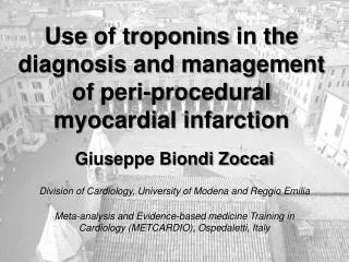
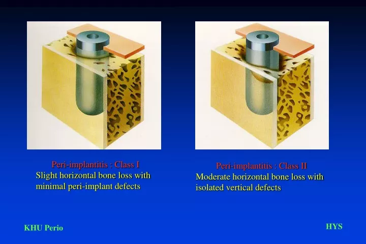
Peri-implantitis : Class II Moderate horizontal bone loss with isolated vertical defects. Peri-implantitis : Class I Slight horizontal bone loss with minimal peri-implant defects. Peri-implantitis : Class III Moderate to advanced horizontal bone loss with broad, circular bony defects.

E N D
Peri-implantitis : Class II Moderate horizontal bone loss with isolated vertical defects Peri-implantitis : Class I Slight horizontal bone loss with minimal peri-implant defects
Peri-implantitis : Class III Moderate to advanced horizontal bone loss with broad, circular bony defects Peri-implantitis Class IV Advanced horizontal bone loss with broad, circumferential vertical defects, as well as loss of oral and/or vestibular bony wall
Single stage implant systems exhibit a coronoapical histologic picture that is comparable to a natural tooth, including an epithelial attachment, a narrow, vasculature -poor zone of connective tissue, and bone. In the connective tissue zone, inner fibers course parallel to the implant surface. The bone-implant is devoid of any connective tissue. With two-stage implants, histology resembling the natural tooth is seldom observed, as the connective tissue is different
Once the inflammatory process spreads beyond the epithelium, the eliciting microorganisms can invade the connective tissue compartment in single-stage implant systems. They advance almost directly into the osseous bed in two-stage systems. Regardless of the type of implant system, however, the host defense capability is limited because it arises from the vasculature elements subjacent to the basal layer of the epithelium
Classification of the degree of resorption of edentulous jaws, according to Lekoholm and Zarb (1985) A.Virtually intact alveolar ridge B. Minor resorption of the alveolar ridge C. Advanced resorption of the alveolar ridge to the base of the dental arch D. Initial resorption of the base of the dental arch E. Extreme risorption of the base of the dental arch.
GTAM- peripheral portion The scanning electron photomicrograph shows the loose texture of the material. This simplifies adaptation to the osseous morphology surrounding the defects. Furthermore, the ingrowth of connective tissue in this area of the membrane should stabilize its position GTAM- central portion The scanning electron photomicrograph reveals the denser texture of the material. This section is considerably stiffer, which helps to create a hollow cavity directly over the defect. The dense structure prevents any penetration by connective tissue cells. Its minimal roughness is reported to favor the proliferation of “pre”-osteoblasts
Classification of bone quality according to Lekoholm and Zarb (1985) Class I : Jaw consists almost exclusively of homogenous compact bone Class II : Thick compact bone surrounds highly trabecular core Class III : Thin cortical bone surrounds highly trabecular core Class IV : Thin cortical bone surrounds loose, spongy core