Download
1 / 31
310 likes | 338 Vues
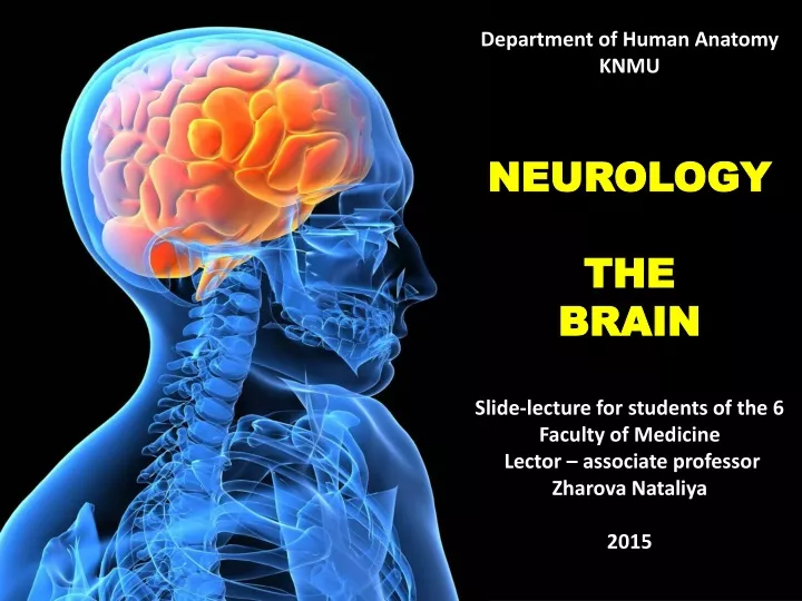
Department of Human Anatomy KNMU NEUROLOGY THE BRAIN Slide-lecture for students of the 6 Faculty of Medicine Lector – associate professor Zharova Nataliya 2015. PLAN The meninges of the brain The brain The telencephalon The texture of the cortex of the cerebral hemispheres

E N D
Department of Human AnatomyKNMUNEUROLOGYTHE BRAINSlide-lecture for students of the 6 Faculty of Medicine Lector – associate professor Zharova Nataliya2015
PLAN Themeningesofthebrain The brain The telencephalon The texture of the cortex of the cerebral hemispheres The grey and white matters The distribution of functions in the cerebral cortex
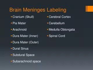
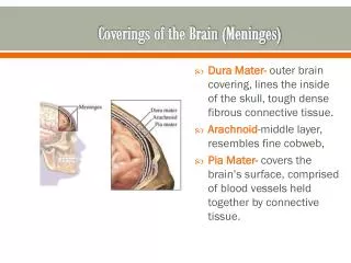
The three meninges of the brain: the dura mater, the arachnoid mater and the pia mater.Spaces: epidural space is absent, subdural, subarachnoid. • The cerebral dura mater a thick whitish connective — tissue membraine is outermost in position. It lines the cranial cavity. It adheres to the internal surfaces of the cranial bones, sending blood vessels and fibrous processes into them. The meningeal layer is falded inwards as four septa that partially divide the cranial cavity into freely communicating spaces in which the subdivisuons of the brain are lodged.
The dura mater consists of the sinuses (10 in number) and the processes (4 in number) The processes: - falx cerebri, between both cewrebral hemispheres, anterriorly attached to crista galli, posteriorly to the terntorium cerebelli - tentorium cerebelli, attached the margins of the transvers sinus of occipital bone, superior margins of the pyramids and temporal bones, Turkish saddle of the sphenoid bone. - falx cerebelli, a small, crescentric fold of dura mater, is below the tentorium cerebelli, projecting forwards into the posterior cerebellar notch. - diaphragma cellae, is a small, circular fold of dura mater, forming a roof to the sella turcica, almost completely covering the hypophysis; a small, central opening in it transmits the infundibulum.
The dura mater contains several reservoirs collecting blood from the brain; these are the sinuses of the dura mater . The sinuses are venous canals devoid of valves and located in the thickness of the dura mater at the attachment of its processes to the skull. The inflexibility of the walls of the venous sinuses provide free drainage of venous activity of the brain. The venous sinuses are as follows. • The transverse, the widest sinus which runs along the posterior margin of the tentorium cerebelli. It descends into the sulcus sinus sigmoidei under the name of the • sigmoid sinus , and at the jugular foramen is continuous with the orifice of the internal jugular vein. • The superior sagittal sinus goes on the superior margin of falx cerebri for the whole length of sulcus sinus sagittalis superioris from crista galli in the internal occipital protuberance. • The occipital sinus is a continuation, as it were, of the superior saggital sinus along the attachment of falx cerebelli to the internal occipital crest and along both margins of the occipital bone. • The straight sinus runs on the line of attachment of falx cerebri to the tentorium cerebelli.
6.The inferior sagittal sinus runs on the free lower margin of falx cerebri and vena cerebri magna. 7. The cavernous sinus is located on the base of the skull lateral to the sella turcica. It is connected with the intercavernous sinuses , passing in front of and behind the hypophyseal fossa. The cavernous sinus receives the superior ophthalmic vein, as well as the inferior end of the 8.sphenoparital sinus running on the margin of the lesser wing of the sphenoid bone. The cavernous sinus is drained of blood by two sinuses locared behind it, namely the 9.superior and 10.inferior petrosal sinuses .
Blood drains from the sinuses into the internal jugular veins.
The sinuses are also connected with the veins of the outer surface of the skull through emissary veins transmitted through openings in the skull bones. The diploic veins and the veins of the spongy substance of the cranial bones also drain into the sinuses of the dura mater, while their other end may be connected with the veins on the external surface of the head.
The arachnoid mater a delicate membrane enveloping the brain, lies between the pia and dural meninges, separated from the dura mater by the subdural space. It is separated from the pia mater be the subarchnoidal space, which is filled with cerebrospinal fluid. The arachnoid surrounds the beginnings of the cranial nerves. • On the upper surface of the brain the arachnoid is thin, but it is thicker on the basal aspect of the brain. Wherever the brain and cranium are not closely adapted, the arachnoid is separated from the pia by wide intervals, named subarachnoid space of which they are merely dilatations. • The arachoid mater has cisterns • - cerebromedullar • - interpeduncular • - chiasmatic • - cistern of the lateral sulcus
The subarachnoid space is connected with the ventricles of the brain by three openings: the median apertura (apertura mediana) in the median plane in the inferior part of the roof of the fourth ventricle and the two lateral apertures (apertura laterales) at the ends of that ventricle's lateral recessus. The arachnoid granulations are small elevations, usually occurring in clusters near the superior sagittal, transverse sinuses. They protrude into the sagittal sinus and its venous lacunae. Fluid injected into the subarachnoid space passes into the granulationes and their villi and thence into the venous sinuses of the dura by osmosis. • The pia mater closely envelopes the brain. It is a vascular membrane, a plexus of minute blood vessels held together by fine loose connective tissue.
The cerebrospinal fluid is a clear fluid. It is secreted into the ventricles of the brain by the choroid plexuses and into the subarachnoid space by plexuses in the fourth ventricle's lateral recesses. From the ventricles it escapes through the apertures of the fourth ventricle into the cerebello — medullary cistern of the subarachnoid space. Intracranially the fluid reaches the arachnoid villi of the superior sagittal sinus, there reentering the bloodstream.
The cerebral arterial circle (of Willis):2 anterior cerebral arteries, 1 anterior communicating artery, 2 posterior communicating arteries, 2 posterior cerebral arteries.
The veins of the brain are divided into superficial and deep veins. The superficial veins collect blood from the cerebral cortex and drain partly into the superior sagittal sinus and partly into the transverse sinus and the sinuses on the base of the skull. • The deep veins receive blood from the central grey nuclei and the ventricles of the brain and merge to form one great cerebral vein. The veins of cerebellum compose the superior and the inferior veins. The superior veins drain blood into the straight sinus and the great cerebral vein. The inferior veins drains into the transverse, sigmoid, and inferior petrosal sinus.
The Brain (encephalon) together with membranes surrounding it is located in the cranial cavity. That is why the convex surface of the brain conforms to the internal concave surface of the arch of the cranium. The inferior surface of the brain is contiguous to the internal base of the skull. It has a complex relief, which conforms to the cranial fossae. The largest parts of the brain are the hemispheres of the telencephalon (cerebral hemispheres), the brainstem and the cerebellum. The diencephalon, the metencephalon (midbrain), the pons and the medulla oblongata form the brainstem. The surface of the cerebral hemispheres is divided into four lobes corresponding to the names of the skull plates that protect them: the frontal lobe, parietal lobe, temporal lobe, and the occipital lobe. In addition to these four lobes, a fifth lobe exists called the insula. This lobe is internal and is not visible from the surface of the brain.
The frontal lobes are sometimes associated with what it means to be human. Absence of the frontal lobes typically results in a person who is deemed emotionally shallow, listless, apathetic, and insensitive to social norms. Control of movement is associated with the frontal lobes via the primary motor cortex located within this lobe. The parietal, temporal, and occipital lobes are specialized for perception. Within the parietal lobe is the primary somatosensory cortex which receives information pertaining to the senses of the body: touch, pressure, temperature, and pain. Visual information is received by the primary visual cortex located within the occipital lobe. Hearing is processed in the primary auditory cortex within the temporal lobe. The central sulcus (fissure of Rolando) divides the frontal lobe from the parietal lobe. The lateral fissure (fissure of Sylvius) separates the temporal lobe from the overlying frontal and parietal lobes. The parieto-occipital fissure separates the parietal and occipital lobes.
The grey matter of hemispheres consist of the cerebral cortex, which is relatively new and basal nuclei –the older structures. The cortex is consist 6 layers: • 1. the molecular layer • 2. the external glandular layer • 3. the external pyramidal layer • 4. the internal glandular layer • 5. the internal pyramidal layer • 6. the multiform layer • The white matter of the hemispheres appear as quite a thick layer between the cerebral cortex and basal nuclei. It consist of nerve fibers arranged into three systems – association (short and long: superior, inferior, cingulum, uncinate fasciculus), commissural, projection pathways.
The corpus callosum is the primary connection between the left and right hemispheres of the cerebral cortex. Connection between the two halves takes place through axons that unite geographically similar regions of the two cerebral cortices. • The basal nuclei: • 1.corpus striatum – 1)caudate nucleus: • head, body, tail; 2)lentiform nucleus: putamen, • globus pallidus; • 2.claustrum • 3.amygdaloid body
The basal ganglia play a central role in a number of neurological conditions, including several movement disorders. The most notable are Parkinson's disease, which involves degeneration of the dopamine-producing cells in the substantia nigra pars compacta, and Huntington's disease, which primarily involves damage to the striatum. Basal ganglia dysfunction is also implicated in some other disorders of behavior control such as Tourette syndrome, hemiballismus, obsessive–compulsive disorder, and Wilson's disease.
The functions of the basal nuclei: • The corpus striatum is the main center of an extrapyramidal system. An extrapyramidal (intrapyramidal) system is a set of the nuclei of the brain and their projective descendent conduction tracts, which make involuntary (automatic) regulation of the impelled acts and muscular tension. • The functions of the extrapyramidal system include maintenance of a pose, organization of the impelled expression of emotions. • All the parts of the extrapyramidal system have diverse communications ensuring realization of the complex impelled actions. • The corpus striatum has some functional differences. The globus pallidus equally with the substantia nigra the red nucleus of the midbrain represents the primary motor center. Involuntary rhythmic motor activity of the newborns is connected with these formations. • The putamen and the caudate nucleus receive proprioceptive impulses from the cerebellum and thalamus. These nuclei coordinate movements on more high level than the corpus striatum. The appearance of the emotional activity of a child is connected with function of the putamen and the caudate nucleus. When these nuclei are injured excessive and involuntary movements and (grimacing, convulsive contraction of muscles) reduction of muscular tension appear. • The claustrum is connected with the olfactory brain, the cortex of the hemisphere of the cerebrum and the thalamus by nervous path ways. The functions of the claustrum are not finally found out.
The corpus callosum is the primary connection between the left and right hemispheres of the cerebral cortex. Connection between the two halves takes place through axons that unite geographically similar regions of the two cerebral cortices. • Under the corpus callosum there is a fornix. • The fornix consists of a body, a column, a crus, a taenia of the fornix. The body of the fornix lies directly under the posterior part of the corpus callosum. On the lateral side of the body of the fornix a vascular plexus of the lateral ventricle is situated. Its epithelium layer is spliced with the fornix. The inferior surface of the body of the fornix adjoins to the thalamus. The right and the left bodies of the fornix are connected by means of cross fibres which have received the name of comissure of the fornix (David's lyra). From ahead the body of the fornix is abruptly bent downwards and continues into the column.
The lateral ventricles. Situated in both hemispheries below the callous body.The parts: - central part, through the parietal lobe. - anterior horn, in frontal lobe. - inferior horn, into temporal lobe. - posterior horn, into occipital lobe.The walls: The superior wall of all horns and central part formed by the fibres of the callous body. The walls of anterior horn: - medial, by septum pellucidum, - lateral and inferior, by the head of the caudate nucleus - superior wall • anterior – genu of callous body. The walls of the central part: - superior wall - inferior, by the body of the caudate nucleus and superior part of thalamus. • medial, by the body of the fornix. The walls of the posterior horn: - superior wall - medial, by projection of the calcarine sulcus • inferior, by trigonum collaterale The walls of the inferior horn: • Superior and lateral walls • medial, by hypocampus • inferior by collateral sulcus.
Choroid plexus of the lateral horn is situated in its central part and in the inferior horn, it is fastened to the taenia of the fornix and the fimbria of the hippocampus. The choroid plexus is presented by a rich in blood capillaries structure projecting into the ventricle's cavity and outwardly.Between lateral ventricles and third ventricle is situated interventricular foramen (Monro)
As the cerebrum is a gross division with many subdivisions and sub-regions, it is important to state that this section lists the functions that the cerebrum as a whole serves. See main articles on cerebral cortex and basal ganglia for more information.
In general terms it is well understood that the left hemisphere controls linguistic consciousness, the right half of the body, talking, reading, writing, spelling, speech communication, verbal intelligence and memories, and information processing in the areas of math, typing, grammar, logic, analytic reasoning, and perception of details. The right hemisphere is associated with 'unconscious' awareness (in the sense it is not linguistically based), perception of faces and patterns, comprehension of body language and social cues, creativity and insight, intuitive reasoning, visual-spatial processing, and holistic comprehension. Communication between the two hemispheres takes place through the corpus callosum, which, by the way, is more fully developed in women than men- likely giving rise to women's intuition.