Download
1 / 17
170 likes | 336 Vues
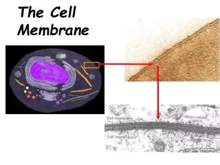
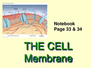
The Cell Membrane. Studying Membranes – The Electron Microscope. Understanding the structure and the function of the phospholipid bilayer cell membrane depended on the invention of the electron microscope.

E N D
Studying Membranes –The Electron Microscope • Understanding the structure and the function of the phospholipid bilayer cell membrane depended on the invention of the electron microscope. • Beams of electrons have a very short wavelength and can resolve cell parts to 0.2 m in size. • U of T graduate students James Hillier and Albert Prebus built the first electron microscope in 1938.
Cell Membrane and Homeostasis • The phospholipidbilayer is responsible for maintaining homeostasis. • Homeostasis is the maintenance of a steady state inside the cell regardless of external changes, to ensure survival. Liver cells - hepatocytes
Cell Membrane Functions • In order to maintain homeostasis the cell must regulate the movement of molecules: • The transport of raw materials into the cell. • The removal of wastes from the cell. • Transfer materials produced in the cell to outside the cell. • Prevent entry of unwanted molecules. • Prevent the escape of needed molecules. • All cell organelles are also surrounded by the same phospholipidbilayer membrane.
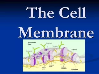
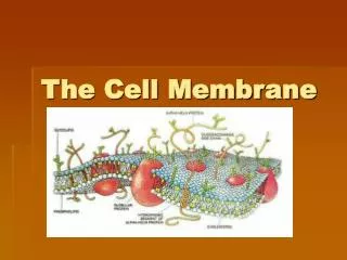
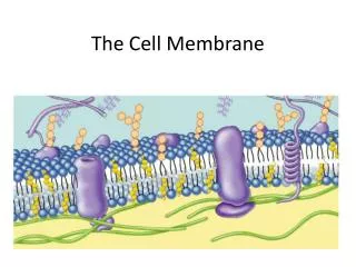
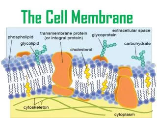
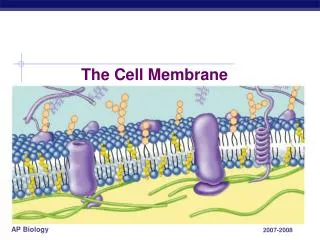
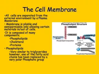
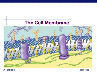
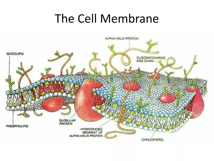
Cell Membrane Structure • There are two phospholipid layers with the hydrophobic ends facing each other on the inside of the bilayer farthest away from the fluids. • Outside the cell is the extracellular (EC) fluid. • Inside the cell is the intracellular (IC) fluid. • The fatty acid bilayer is selectively permeable.
Phospholipids in bilayer Intracellular Fluid Hydrophilic head Hydrophobic tail Extracellular Fluid
The Membrane is Flexible and Fluid More Viscous More Fluid Saturated hydrocarbon tails Unsaturated hydrocarbon tails with kinks When the membrane is colder it is less fluid and a warmer membrane is more fluid. This is due to cholesterol molecules in the membrane.
The Membrane is Dynamic Lateral movement (~107 times per second) Flip-flop (~ once per month) Movement of phospholipids
Fluid Mosaic Model • Our current understanding of cell membranes is called the Fluid Mosaic Model. • There are many molecules within the membrane and other molecules connected to the membrane. • Some small non-polar molecules, like oxygen and carbon dioxide, can pass through the hydrophobic part of the cell membrane. • Even water, which is slightly polar, can pass through the hydrophobic cell membrane at a low rate.
Cell Membrane Proteins • Carrier proteins are transmembrane proteins that help move neutral water soluble molecules, like glucose, from outside to inside the cell. • Channel proteins are also transmembrane and allow ions to pass through the membrane. • Aquaporins are special proteins that help water pass through the cell membrane.
Cell Membrane Carbohydrates • The lipids and the proteins often have unique carbohydrates attached to them. • These carbohydrates are used as recognition sites for molecules attaching to the cell membrane. • Glycoproteins also serve as markers for anchors to other internal structures like the cytoskeleton. • Glycolipids also serve as recognition sites for other molecules as well as joining cells to form tissues.
Videos and Animations • http://www.youtube.com/watch?v=Qqsf_UJcfBc • http://www.youtube.com/watch?v=moPJkCbKjBs&feature=related • http://www.youtube.com/watch?v=Rl5EmUQdkuI&feature=related • http://www.teachersdomain.org/resource/tdc02.sci.life.cell.membraneweb • (also includes molecule movement) • http://www.teachersdomain.org/resource/tdc02.sci.life.cell.membraneweb