Download
1 / 42
420 likes | 525 Vues
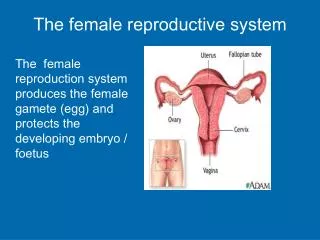
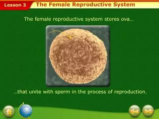
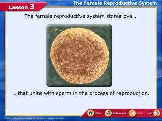
The Female Reproductive System. Exercise 31 A&P 233. Female Reproduction. Unlike males, who are able to produce sperm cells throughout their reproductive lives, females produce a finite number of egg cells.

E N D
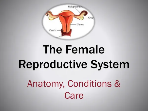
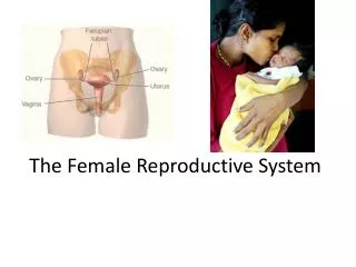
The Female Reproductive System Exercise 31 A&P 233
Female Reproduction • Unlike males, who are able to produce sperm cells throughout their reproductive lives, females produce a finite number of egg cells. • During early fetal development germ cells migrate into the ovaries and differentiate into oogonia
Oogonia • The oogonia divide by mitosis for the next few months and some differentiate into primary oocytes. • By fifth month there are about 7 million primary oocytes, but most will degenerate during the next 2 months
Oogonia • Those that remain will be surrounded by a single layer of squamous epithelial cells (follicle cells) called a primordial follicle. • Degeneration of primary oocytes continues. • At birth =1million primordial follicles • At puberty 400,000 remain • Only 400-500 will reach maturity
Ovarian Cycle • Monthly changes that occur in the ovary during a woman’s reproductive life. • Each month FSH stimulates primordial follicles to grow and mature (follicular phase) • Ovulation- release of the egg (LH) • Luteal phase the corpus luteum produces progesterone that maintains uterine walls If fertilization does not occur, the corpus luteum degenerates, within 2 weeks into a mass of scar tissue called the corpus albicans
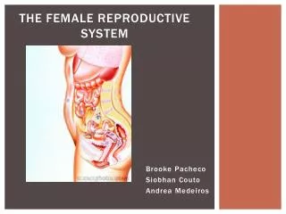
Gross Anatomy • The ovaries are solid, ovoid structures, about 2 cm in length and 1 cm in width. • Like the testes, they develop from embryonic tissue along the posterior abdominal wall, near the kidneys. • Accessory organs include the uterine tubes, uterus, and vagina.
Uterine Tubes (Fallopian Tubes) • Receive the ovulated oocyte and provide a site for fertilization • Empty into the superolateral region of the uterus via the isthmus • Expand distally around the ovary forming the ampulla • The ampulla ends in the funnel-shaped, ciliated infundibulum containing fingerlike projections called fimbriae
Uterine Tubes (Fallopian Tubes) • Function: events occurring in the uterine tube • Fimbriae sweep oocyte into tube, cilia & peristalsis move it along, sperm reaches oocyte in ampulla, fertilization occurs within 24 hours after ovulation & zygote reaches uterus about 7 days after ovulation
Fallopian Tube Histology Cilia sweep egg/zygote toward the uterus
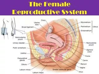
Uterus • Hollow, thick-walled organ located in the pelvis anterior to the rectum and posterosuperior to the bladder • Body: Major portion of the uterus • Fundus: Rounded region superior to the entrance of the uterine tubes • Isthmus: Narrowed region between the body and cervix
Uterine Histology • Endometrium • Simple columnar epithelium • Stroma of connective tissue and endometrial glands • Stratum functionalis: Shed during menstruation • Stratum basalis: Replaces stratum functionalis each month • Myometrium • 3 layers of smooth muscle • Perimetrium • Visceral peritoneum
Endometrium Simple columnar epithelium Endometrial glands
Endometrium • Proliferative phase: glands and blood vessels scattered throughout the functional zone with little or no branching. • New glands form and endometrium thickens. • Secretory phase: glands are enlarged and have branches. Preparing the endometrium for implantation • If no implantation then endometrium breaks down and menstruation begins.
Cervix • Narrow lower neck of the uterus which projects into the vagina inferiorly • Cervical canal – cavity of the cervix that communicates with: • The vagina via the external os • The uterine body via the internal os • Cervical glands secrete mucus that covers the external os and blocks sperm entry except during midcycle
Endocervical canal Fornix
Vagina • Thin-walled tube lying between the bladder and the rectum, extending from the cervix to the exterior of the body • Wall consists of three coats: fibroelastic adventitia, smooth muscle muscularis, and a stratified squamous mucosa • Mucosa near the vaginal orifice forms an incomplete partition called the hymen • Vaginal fornix: upper end of the vagina surrounding the cervix
Female External Genitalia • Mons pubis: fatty pad over the pubic symphysis • Labia majora & minora: folds of skin encircling vestibule where find urethral and vaginal openings • Clitoris: small mass of erectile tissue • Bulb of vestibule: masses of erectile tissue just deep to the labia on either side of the vaginal orifice • Perineum: Area between the vagina and anus
Female External Genitalia Perineum
Bartholin’s Glands (aka: Vestibular Glands) • The Bartholin's glands are located on each side of the vaginal opening. • They secrete fluid that helps lubricate the vagina. • Sometimes the ducts of these glands become obstructed. • Fluid backs up into the gland and causes swelling (Bartholin's cyst)
Mammary Glands • Modified sweat glands that produce milk (lactation) • Amount of adipose determines size of breast • Milk-secreting glands open by lactiferous ducts at the nipple • Areola is pigmented area around nipple • Suspensory ligaments suspend breast from deep fascia of pectoral muscles (aging & Cooper’s droop) • Mammary line is a thickened ridge of embryonic tiwwue that extends from the axilla to the groin.
Breast • Prolactin from the pituitary gland stimulates the synthesis of milk • Oxytocin from the posterior pituitary gland stimulates milk ejection
Lymphatic Drainage • Lymph nodes draining the breast are located in the axilla.
Oogenesis: Before birth • During fetal development, oogonia (stem cells) divide by mitosis to make primary oocytes • Primary oocytes begin meiosis and stop in prophase I until puberty • Primordial follicles: Support cells that surround the oocyte in the ovary • 2 million present at birth • 400,000 remain at puberty
Oogenesis: After Puberty • Each month, hormones cause several follicles to develop, which triggers the primary oocyte to resume meiosis I • Polar bodies: When the cell divides, all the cytoplasm and organelles stay with one of the new cells, the other cell is just DNA, and is called a polar body and is discarded • Secondary oocyte: The stage at which ovulation occurs.
Oogenesis: After Puberty • The secondary oocyte begins meiosis II, but stops in metaphase II • The secondary oocyte is ovulated • Meiosis II is completed only if it is fertilized.
Life History of Oogonia • As a fetus, oogonia divide to produce millions by mitosis but most degenerate (atresia) • Some develop into primary oocytes & stop in prophase stage of meiosis I • 200,000 to 2 million present at birth • 40,000 remain at puberty but only 400 mature during a woman’s life • Each month, hormones cause meiosis I to resume in several follicles so that meiosis II is reached by ovulation • Penetration by the sperm causes the final stages of meiosis to occur
Ovaries • Each follicle consists of an immature egg called an oocyte • Cells around the oocyte are called: • Follicle cells (one cell layer thick) • Stimulated to mature by FSH from the pituitary gland • Granulosa cells (when more than one layer is present) • Thecal cells: Cells in the ovarian stroma • Thecal & granulosa cells work together to produce estrogen • A protective layer of glycoprotein forms around the egg called the zona pellucida
Follicle Development • Primordial follicle: one layer of squamous-like follicle cells surrounds the oocyte • Primary follicle: two or more layers of cuboidal granulosa cells enclose the oocyte • Secondary follicle: has a fluid-filled space between granulosa cells that coalesces to form a central antrum • Graafian follicle: secondary follicle at its most mature stage that bulges from the surface of the ovary • Corpus luteum : ruptured follicle after ovulation
Primary Follicle 1° Oocyte(arrested in prophase I) Nucleus Primordial follicle Zona pellucida Thecal cells Granulosa cells
Secondary Follicle Fluid-filled antrum
Graafian Follicle Fluid filled antrum Oocyte 2° Granulosa cells Stalk Corona radiata Zona pellucida
Corpus luteum • After ovulation, the remains of the follicle are transformed into a structure called the corpus luteum. • If a pregnancy occurs, it produces progesterone to maintain the wall of the uterus during the early period of development.
Corpus albicans • If fertilization does not occur, the corpus luteum will begin to break down about 2 weeks after ovulation. • Degeneration occurs when fibroblasts enter the corpus luteum and a clump of scar tissue forms called the corpus albicans.
Today’s Activities • View female reproductive organs on the models • View slides of ovaries, fallopian tubes, uterus-proliferative, secretive, menstrual