Download
1 / 1
10 likes | 84 Vues
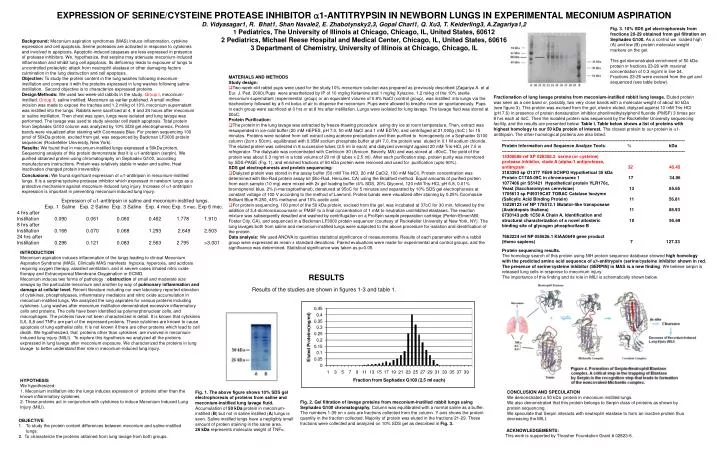
Fig. 3. 10% SDS gel electrophoresis from fractions 20-29 obtained from gel filtration on Sephadex G100. As a control we loaded high (A) and low (B) protein molecular weight markers on the gel.

E N D
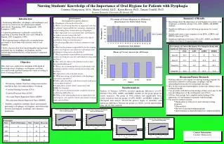
Fig. 3. 10% SDS gel electrophoresis from fractions 20-29 obtained from gel filtration on Sephadex G100. As a control we loaded high (A) and low (B) protein molecular weight markers on the gel. This gel demonstrated enrichment of 50 kDa protein in fractions 23-26 with maximal concentration of 0.3 mg/ml in line 24. Fractions 23-26 were excised from the gel and sequenced (see table below). • MATERIALS AND METHODS • Study design: • Two-week-old rabbit pups were used for the study.10% meconium solution was prepared as previously described (Zagariya A. et al Eur. J. Ped. 2000).Pups were anesthetized by IP of 10 mg/kg Ketamine and 1 mg/kg Xylazine. 1.2 ml/kg of the 10% sterile meconium supernatant (experimental group) or an equivalent volume of 0.9% NaCl (control group), was instilled into lungs via the tracheotomy followed by a 5 ml bolus of air to disperse the meconium.Pups were allowed to breathe room air spontaneously. Pups in each group were sacrificed at 0 hrs or at 8 hrs after instillation. Lungs were isolated for lung lavage. The lavage fluid was stored at 20oC. • Protein Purification: • The protein in the lung lavage was extracted by freeze-thawing procedure using dry ice at room temperature. Then, extract was resuspended in ice-cold buffer (20 mM HEPES, pH 7.0, 50 mM NaCl and 1 mM EDTA), and centrifuged at 21,000g (4oC ) for 15 minutes. Proteins were isolated from cell extract using acetone precipitation and then purified to homogeneity on a Sephadex G100 column (2cm x 50cm), equilibrated with 0.05M sodium phosphate buffer at pH 7.0, the protein was eluted in 0.1 M sodium chloride. The eluted protein was collected in 8 successive tubes (2.5 ml in each) and dialyzed overnight against 20 mM Tris-HCl, pH 7.5 in refrigerator. The dialysate was concentrated with Centricon-30 (Amicon, Beverly, MA) and was stored at –80oC. The yield of the protein was about 0.3 mg/ml in a total volume of 20 ml (8 tubes x 2.5 ml). After each purification step, protein purity was monitored by SDS-PAGE (Fig. 1), and enriched fractions of 50 kDa protein were removed and used for purification (upto 90%). • SDS gel electrophoresis and protein sequencing: • Dialyzed protein was stored in the assay buffer (50 mM Tris-HCl, 20 mM CaCl2, 100 mM NaCl). Protein concentration was determined with Bio-Rad protein assay kit (Bio-Rad, Hercules, CA) using the Bradford method. Equal amounts of purified protein from each sample (10 mg) were mixed with 2x gel loading buffer (4% SDS, 20% Glycerol, 120 mM Tris-HCl, pH 6.8, 0.01% bromophenol blue, 2% b-mercaptoethanol), denatured at 95oC for 5 minutes and separated by 10% SDS gel electrophoresis at constant voltage of 100 V according to the method of Laemmli. Protein bands were visualized after staining by 0.25% Coomassie Brilliant Blue R-250, 45% methanol and 10% acetic acid. • For protein sequencing, 100 pmol of the 50 kDa protein, excised from the gel, was incubated at 37oC for 30 min, followed by the addition of 3,4-dichloroisocoumarin or PMSF to a final concentration of 1 mM to neutralize uninhibited elastases. The reaction mixture was subsequently desalted and washed by centrifugation on a ProSpin sample preparation cartridge (Perkin-Elmer/ABI; Foster City, CA), and sequenced in a Beckman LF3000 protein sequencer (courtesy of Rockefeller University at New York, NY). The lung lavages both from saline and meconium-instilled lungs were subjected to the above procedure for isolation and identification of the protein. • Data analysis: We used ANOVA to quantities statistical significance of measurements. Results of each parameter within a rabbit group were expressed as mean ± standard deviations. Paired evaluations were made for experimental and control groups, and the significance was determined. Statistical significance was taken as p<0.05. EXPRESSION OF SERINE/CYSTEINE PROTEASE INHIBITOR a1-ANTITRYPSIN IN NEWBORN LUNGS IN EXPERIMENTAL MECONIUM ASPIRATIOND. Vidyasagar1, R. Bhat1, Shan Navale2, E. Zhabotynsky2,3, Gopal Chari1, Q. Xu3, T. Keiderling3, A.Zagariya1,21 Pediatrics, The University of Illinois at Chicago, Chicago, IL, United States, 606122 Pediatrics, Michael Reese Hospital and Medical Center, Chicago, IL, United States, 606163 Department of Chemistry, University of Illinois at Chicago, Chicago, IL Fractionation of lung lavage proteins from meconium-instilled rabbit lung lavage. Eluted protein was seen as a one band or, possibly, two very close bands with a molecular weight of about 50 kDa (see figure 3). This protein was excised from the gel, electro eluted, dialyzed against 10 mM Tris-HCl (pH 7.6) in presence of protein denaturation inhibitor phenilmethylsylphonil fluoride (PMSF) 3 times per 8 hrs each at 4oC. Then the isolated protein was sequenced by the Rockefeller University sequencing facility, and the results are presented below. Table I. Table below shows a list of proteins with highest homology to our 50 kDa protein of interest. The closest protein to our protein is a1-antitripsin. The other homological proteins are also listed. --------------------------------------------------------------------------------------------------------------------------- Protein Information and Sequence Analyze Tools: % kDa --------------------------------------------------------------------------------------------------------------------------- 15300686 ref XP 028358.2 serine (or cysteine) protease inhibitor, clade A (alpha-1 antiprotease, antitrypsin 32 40.45 3183393 sp O1377 YE99 SCHPO Hypothetical 35 kDa Protein C17A5.09C in chromosome 1 17 34.96 1077406 pir S51421 Hypothetical protein YLR176c, Yeast (Saccharomyces cerevisiae) 13 85.65 1705613 sp P49319CAT TOBAC Catalase Isozyme (Salicylic Acid Binding Protein) 11 56.81 15228123 ref NP 178512.1 Mutator-like transposase (Arabidopsis thaliana) 11 88.93 6730143 pdb 1C50 A Chain A, Identification and structural characterization of a novel allosteric 10 95.69 binding site of glycogen phosphorilase B 7662224 ref NP 055626.1 KIAA0649 gene product (Homo sapiens) 7 127.33 Expression of a1-antitripsin in saline and meconium-instilled lungs. Exp. 1 Saline Exp. 2 Saline Exp. 3 Saline Exp. 4 mec Exp. 5 mec. Exp 6 mec. 4 hrs after Instillation 0.090 0.061 0.060 0.462 1.778 1.910 8 hrs after Instillation 0.168 0.070 0.068 1.293 2.649 2.503 24 hrs after Instillation 0.296 0.121 0.083 2.563 2.795 >3.001 Background: Meconium aspiration syndromes (MAS) induce inflammation, cytokine expression and cell apoptosis. Serine proteases are activated in response to cytokines and involved in apoptosis. Apoptotic-induced caspases are less expressed in presence of protease inhibitors. We, hypothesize, that serpine may attenuate meconium-induced inflammation and inhibit lung cell apoptosis. Its deficiency leads to exposure of lungs to uncontrolled proteolytic attack from neutrophil elastase or other damaging factors culmination in the lung destruction and cell apoptosis. Objective: To study the protein content in the lung washes following meconium-instillation and compare it with the proteins expressed in lung washes following saline-instillation. Second objective is to characterize expressed proteins.Design/Methods: We used two-week-old rabbits in the study: Group I, meconium-instilled. Group II, saline instilled. Meconium as earlier published. A small midline incision was made to expose the trachea and 1.2 ml/kg of 10% meconium supernatant was instilled into the lungs. Rabbits were sacrificed at 4, 8 and 24 hours after meconium or saline instillation. Then chest was open, lungs were isolated and lung lavage was performed. The lavage was used to study alveolar cell death apoptosis. Total protein from Sephadex G100 column was analyzed by 10% SDS gel electrophoresis. Protein bands were visualized after staining with Coomassie Blue. For protein sequencing 100 pmol of 50kDa protein, excited from gel, was sequenced by Backman LF3000 protein sequencer (Rockefeller University, New York). Results: We found that in meconium-instilled lungs expressed a 50kDa protein, Sequencing analysis of this protein demonstrate that it is a1-antitripsin (serpin). We purified obtained protein using chromatography on Sephadex G100, according manufacturers instructions. Protein was relatively stable in water and saline. Heat inactivation changed protein irreversibly. Conclusions: We found significant expression of a1-antitripsin in meconium-instilled lungs. It is a serpine/sycteine protease inhibitor which expressed in newborn lungs as a protective mechanism against meconium-induced lung injury. Increase of a1-antitripsin expression is important in preventing meconium induced lung injury. INTRODUCTION Meconium aspiration induces inflammation of the lungs leading to clinical Meconium Aspiration Syndrome (MAS). Clinically MAS manifests hypoxia, hyperoxia, and acidosis requiring oxygen therapy, assisted ventilation, and in severe cases inhaled nitric oxide therapy and Extracorporeal Membrane Oxygenation or ECMO. Meconium induces two forms of pathology : obstruction of small and moderate size airways by the particulate meconium and another by way of pulmonary inflammation and damage at cellular level. Recent literature including our own laboratory reported elevation of cytokines, phospholipases, inflammatory mediators and nitric oxide accumulation in meconium-instilled lungs. We analyzed the lung aspirates for various proteins including cytokines. Lung washes after meconium instillation demonstrated excessive inflammatory cells and proteins. The cells have been identified as polymorphonuclear cells, and macrophages. The proteins have not been characterized in detail. It is known that cytokines IL6, IL8 and TNFa are part of the expressed proteins. These cytokines are known to cause apoptosis of lung epithelial cells. It is not known if there are other proteins which lead to cell death. We hypothesized, that proteins other than cytokines are involved in meconium-induced lung injury (MILI). To explore this hypothesis we analyzed all the proteins expressed in lung lavage after meconium exposure. We characterized the proteins in lung lavage to better understand their role in meconium-induced lung injury. Protein sequencing results. The homology search of this protein using NIH protein sequence database showed high homology with the predicted amino acid sequence of a1-antitrypsin (serine/cysteine inhibitor shown in red. The presence of serine/cysteine inhibitor (SERPIN) in MAS is a new finding. We believe serpin is released lung cells in response to meconium injury. The importance of this finding and its role in MILI is schematically shown below. RESULTS Results of the studies are shown in figures 1-3 and table 1. HYPOTHESIS We hypothesized: 1. Meconium instillation into the lungs induces expression of proteins other than the known inflammatory cytokines. 2. These proteins act in conjunction with cytokines to induce Meconium Induced Lung Injury (MILI). CONCLUSION AND SPECULATION We demonstrated a 50 kDa protein in meconium instilled lungs. We also demonstrated that this protein belongs to Serpin class of proteins as shown by protein sequencing. We speculate that Serpin interacts with neutrophil elastase to form an inactive protein thus decreasing the MILI. Fig. 1. The above figure shows 10% SDS gel electrophoresis of proteins from saline and meconium-instilled lung lavage fluid. Accumulation of 50 kDa protein in meconium-instilled (B) but not in saline instilled (A) lungs is seen. Saline instilled lungs have a negligibly small amount of protein staining in the same area. 26 kDa represents molecular weight of TNFa. Fig. 2. Gel filtration of lavage proteins from meconium-instilled rabbit lungs using Sephadex G100 chromatography. Column was equilibrated with a normal saline as a buffer. The numbers 1-39 on x axis are fractions collected from the column. Y axis shows the protein quantity in the fraction collected. Majority of protein was eluted in the fractions 21-29. These fractions were collected and analyzed on 10% SDS gel as described in Fig. 3. OBJECTIVE 1. To study the protein content differences between meconium and saline-instilled lungs. 2. To characterize the proteins obtained from lung lavage from both groups. ACKNOWLEDGEMENTS: This work is supported by Thrasher Foundation Grant # 02823-6.