Download
1 / 43
430 likes | 446 Vues
Cerevrovascular disease. Dr. Basu MD. Topic. Cerebral (intracranial) hemorrhage Types Etiology Morphology Clinical features. Intracranial hemorrhage: types. Non traumatic [ Spontaneous hemorrhage] Intracerebral ( Primary Brain Parenchymal Hemorrhage ) Sub-arachnoid Hemorrhage

E N D
Cerevrovascular disease Dr. Basu MD
Topic • Cerebral (intracranial) hemorrhage • Types • Etiology • Morphology • Clinical features
Intracranial hemorrhage: types • Non traumatic [Spontaneous hemorrhage] • Intracerebral (Primary Brain Parenchymal Hemorrhage) • Sub-arachnoid Hemorrhage • Mixed Hemorrhage • Traumatic
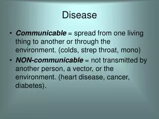
Spontaneous hemorrhage:Etiology A . Intracerebral (intraparenchymal) - predominantly, hypertensive. B. Subarachnoid - predominantly, aneurysmal. C. Mixed intracerebral and subarachnoid - usually associated with rupture of arteriovenous malformations.
Spontaneous Intracerebral / intraparenchymal hemorrhage AGE : MIDDLE TO LATE ADULT LIFE PICK AGE : 60 YEARS Cause: 1.Acute and chronic raise of blood pressure. 2. Rupture of Charcot – Bouchard microaneurysm 50 % of all hemorrhage
Risk factors: It can develop in an ischemic Infract particularly if reperfusion occur. If the patient is on anticoagulant therapy In case of Amyloid angiopathy. Location: Putamen – 60% Thalamus Pons Spontaneous Intracerebral / intraparenchymal hemorrhage
Hemorrhages involving the basal ganglia area ; Hypertensive hemorrhage
Rupture of Charcot – Bouchard microaneurysm • IT IS A FEATURE OF CHRONIC HYPERTANSION • Involve a vessels less than 300micrometer in Diameter. • SITE: BASAL GANGLIA
Complication of Intracranial hemorrhage • Mass effect can cause Herniation • Secondary Brain stem or Duret hemorrhage
Clinical Features : Hypertensive hemorrhage • Sudden loss of consciousness • Vomiting, Headache [ increased intracranial pressure ] • EFFECT OF BRAIN STEM COMPRESSION • IRREGULAR RESPIRATION • PERIOD OF APNEA [ CHEYNE STROKE BREATHING] • DILATED NONRESPONSIVE PUPIL • SPASTICITY
SUBARACHONOID HAEMORRHAGE • Etiology: • RUPTURE OF SACCULAR Aneurysm ( Berry aneurysm ) • Size: 6-10 mm.
Berry aneurysms arise in a weak (ness) point in the arterial wall (media). Disease associated: Marfan syndrome, Ehlar Danlos, Adult polycystic kidney disease. Berry aneurysm Angiogram
Morphology If less than 3 mm in diameter = asymptomatic. 30 % Cases multiple. CSF : will show blood.
Clinical features : Subarachnoid Hemorrhage • Women are effected more than male. • Giant aneurysm > 25 mm in diameter. • It gradually enlarges from child hood, become large enough to produce symptoms in adulthood by sudden increased blood pressure.
Clinical Signs : In subarachnoid Hemorrhage. • Headache, come , vomiting is present, herniation, acute Hydrocephalous. Additional features: • EVIDENCE OF MENINGIAL IRITATION IS PRESENT • 1. NECK RIGIDITY • 2. BLOODY CSF
Mixed intracerebral and subarachnoid hemorrhage Cause: Vascular malformations. This are 4 TYPES • ARTERIOVANOUS MALFORMATION • CAPILLARY TELANGECTASIS • VENOUS ANGIOMA • CAVERNOUS ANGIOMA
ARTERIOVANOUS MALFORMATION • Common in the cerebral hemispheres. Males are affected twice as often as females. • AGE = 10 – 30
“HAPHAZARDLY ARRANGED BLOOD VESSELS, CONTAINING ARTERIES AND VEINS AND TRANSITIONAL FORMS OF THE VESSELS”. Microscopy
Classification according to the anatomical Location of the Hemorrhage. THREE TYPES 1. Epidural Hematoma 2. Sub-dural Hematoma. 3. Traumatic parenchymal Injuries.
Epidural Hematoma Cause Rupture of Meningeal Artery on the occasion of a skull fracture. X- ray: show evidence of lateral skull area fracture.
Clinical Effect : Mass Effect • If not immediately drained: • It will cause the following effects :- 1. Uncal and Tonsillar Herniation 2. Brain Stem compression and DEATH
Sub-Dural Hematoma • Cause: Rupture of the Bridging Veins
CT Showing the Hematoma CAUSE: Key: Small brain big skull. More brain mobility. RAPID CANGE IN THE HEAD Velocity. Types: Acute Subdural Hematoma (child) Chronic subdural hematoma ( adult)
Acute Subdural Hematoma • Seen in infants • Cause: • More spacer in cranium, less brain matter • Excess brain mobility • Hemorrhage due to minor trauma.
CHRONIC SUBDURAL HEMATOMA • Relative slow progress, contain venous blood. • Associated with brain atrophy, which gives ‘brain’ more mobility. • This make the vein more vulnerable to trauma.
CHRONIC SUBDURAL HEMATOMA : CLINICAL PICTURE HEAD Injury, or, Minor trauma 2 weeks/2 months Onset of Headache, dizziness, consciousness disturbance.
Traumatic Parenchymal Injuries • Concussion • Contusion and Laceration • Intracerebral hemorrhage • Generalized brain edema.
Concussion • Definition • Concussion may result when the head strikes against an object or is struck by an object. • Concussions may produce unconsciousness or bleeding in or around the brain.
Concussion • It is associated with minimum morphological change. • Unconsciousness is due to the injury to the Reticular activating System.
Contusion and Laceration • Contusion = Injury to theSuperficial Brain parenchyma due to the Blunt trauma. • Laceration = CONTUSION +TEAR OF THE SUPERFICIAL LAYERS OF THE BRAIN.
Coup Contusion Hit on Immobile Head Injury Directly beneath the area of blunt force: no skull fracture
Contrecoup contusion INJURY In fall if the occipital area strikes the floor Injury in the Frontal and temporal poles area in a mobile head.
Laceration • CONTUSION +TEAR OF THE SUPERFICIAL LAYERS OF THE BRAIN.
Traumatic Intracerebral hemorrhage • Multiple often associate with contusion and edema of the Brain.
Diffuse axonal injury • Injury to white matter • Damage to axon at node of Ranvier. • Common sites: • Corpus callosum, periventricular area, hippocampus. • Clinical: coma after trauma without evicence of parenchymal injury. • Histology: axonal swelling.