Download
1 / 32
320 likes | 608 Vues
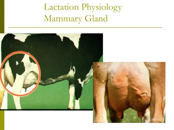
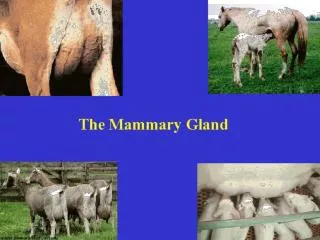
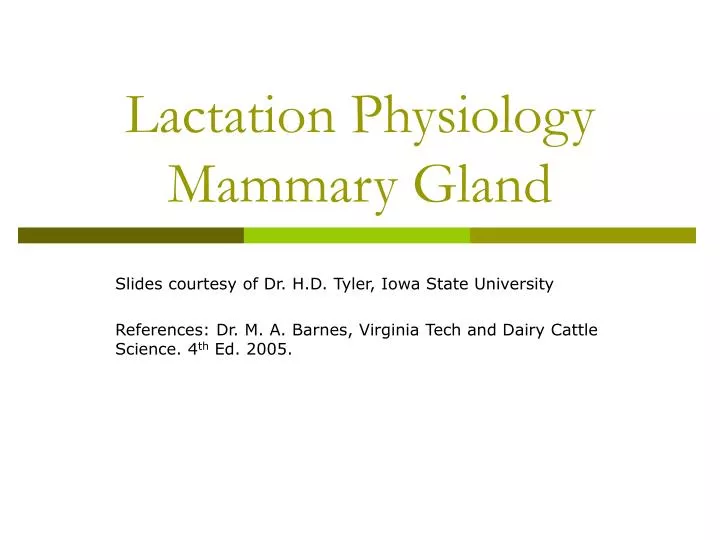
Lactation Physiology Mammary Gland. Slides courtesy of Dr. H.D. Tyler, Iowa State University References: Dr. M. A. Barnes, Virginia Tech and Dairy Cattle Science. 4 th Ed. 2005. The mammary gland nourishes the neonate. Exocrine gland; common to all mammals Function: nourish the neonate

E N D
Lactation PhysiologyMammary Gland Slides courtesy of Dr. H.D. Tyler, Iowa State University References: Dr. M. A. Barnes, Virginia Tech and Dairy Cattle Science. 4th Ed. 2005.
The mammary gland nourishes the neonate • Exocrine gland; common to all mammals • Function: nourish the neonate • Food source: fat, protein, sugar (CHO), vitamins, minerals, water • Protection: immunoglobulins (first Ab protection; absorbed via intestinal tract
The mammary gland is part the reproductive system • The mammary gland is loosely considered part of the reproductive system: • Serves a “reproductive function”; nourishment of the neonate = survival of species. • Relies on same endocrine (hormonal) support for development and function. Example: gonadal steroids, prolactin, etc.
Mammary Gland Structure • Udder consists of four separate glands • A teat hangs from each quarter • Bottom of teat closed by sphincter muscle known as streak canal • Can have extra nonfunctional teats • Called supernumerary teats • Removed when calf is young • Conformation of teats • Vary in shape from cylindrical to conical • Rear teats are usually shorter • Each teat has one streak canal • Teats should be moderately sized and located centrally on each quarter • Sphincter in each teat should be tight enough to prevent leakage • Teats are hairless
Mammary Gland Structure • Support system = Stroma (connective tissue) • Glandular; secreting tissue = Parenchyma • Alveoli- secreting epithelial cells • Duct system- lined by epithelial cells • Lobules & lobes- clusters of alveolar tissue supported by connective tissue
Separate Mammary Glands-Quarters 40% 60%
Mammary Gland Structure/Suspension • Intermammary groove separates left and right halves of the udder • Udder can weigh anywhere from 7 to 165 pounds • May support up to 80 pounds of milk • Rear quarters secrete 60% of the milk • Udder continues to grow in size until cow is 6 years of age • Well attached udder fits snugly against the abdominal wall in front and on the sides • Extends high between thighs in rear • 3 major supporting structures • Skin • Median suspensory ligament • Lateral suspensory ligament
Mammary Gland Suspension • Skin • Minor role in support • Median suspensory ligament • Separates right and left halves of udder • Connects udder to abdominal wall • Lamellae • Elastic tissue which responds to weight of milk in udder • Lateral suspensory ligament • Inflexible • Surround the outer wall of udder • Attached to prepubic and subpubic tendons • Intermammary groove formed where lateral suspensory ligament and median suspensory ligament meets
Fig 29-3. An illustrated view of the ligaments that permit udder suspension (Courtesy of Iowa State University)
Mammary Gland Support Medial suspensory ligament
Internal Anatomy • Streak canal • Functions to keep milk in udder and bacteria out of udder • Teat cistern • Duct in teat with capacity of 30-45 milliliters • Separated from streak canal by folds of tissue called Furstenberg’s rosettes • Gland cistern • Separated from teat cistern by the cricoid fold • Holds up to 400 milliliters of milk • Collecting area for the mammary ducts • From this branches the mammary ducts
Fig 29-4. A dissected mammary gland showing the gland cistern, teat cistern and streak canal (Courtesy of Mark Kirkpatrick)
Alveoli and Duct System • Alveoli is the basic milk producing unit • Small bulb-shaped structure with hollow center • Lined with epithelial cells that secrete milk • Each cubic inch of udder tissue contains 1 million alveoli • Each alveoli surrounded by network of capillaries and myoepithelial cell • Contraction of myoepithelial cell stimulates milk ejection • Groups of alveoli empty into a duct forming a unit called a lobule • Several lobules create a lobe • Ducts of lobe empty into a galatophore, which empties into the gland cistern • Ducts provide storage area for milk and a means for transporting it outside • Lined by two layers of epithelium • Myoepithelial cells are arranged in longitudinal pattern • Shorten to increase diameter to facilitate flow of milk
Alveolar Products • Alveolus: • basic secretory unit; lined by epithelial cells which synthesize and/or secrete: • lipid - triglycerides & free fatty acids (FFA) • protein - caseins • lactose - disaccharide; major CHO; osmoreactive molecule (draws water) • minerals & vitamins - Ca, P, K; Vits. A, B, C, D • water
Alveolar Structure • Alveolar components & function: • epithelial cells - milk synthesis & secretion • lumen - collect milk components & water • myoepithelial cells - milk ejection • basement membrane - selective transfer • terminal duct - milk transport out of alveoli • capillary system - supply milk precursors and deliver hormones
Mammary Cell Function • Alveolar milk component synthesis: • RER > lipid, caseins • Golgi apparatus > lactose (also packages lactose, caseins, minerals, water)
Circulation • One gallon of milk requires 400 gallons of blood being passed through udder • Ratio may increase in low producing cows • Blood enters the udder through external pudic arteries • Blood exiting udder from veins at the base of udder blood can travel through two routes • Via external pudic veins • Via subcutaneous abdominal veins
Fig 29-6. Blood flow to and from the mammary gland determines milk producing capability of the cow (Courtesy of Iowa State University)
Mammary Venous Circle Cranial Mammary Vein
Lymphatic System • Lymph is clear, colorless • contains less protein than blood plasma • contains high [ ] of lymphocytes (WBC’s) which play a role in immune defense • contains few RBC’s • carries glucose, salts, fat (chylomicra from intestine) • dissipates heat • carrier of fibrinogen (clotting protein)
Lymphatic System • Movement of lymph is passive: • lymph moves through vessels by: • muscle movement (exercise, etc.) • breathing • heart beat • tissue massage
Lymphatic System • Helps regulate proper fluid balance within udder and combat infection • Fluid drained from tissue only travels away from udder • Blood capillary pressure • Contraction of muscles surrounding the lymph vessels • Valves that prevent backflow of lymph • Mechanical action of breathing • Lymph travels from udder to the thoracic duct and empties into blood system • Flow rates of lymph depend on physiological status of the cow
Lymphatic System • Fluid enters the lymph system through open-ended vessels called lacteals
Lymphatic System- Edema • Edema: • low pressure, passive system fed by a high pressure vascular system! • this situation results in pooling of interstitial fluid if evacuation of lymph is impaired Example: tissue trauma; increased mammary blood flow at parturition
Alleviating Mammary Edema • Preparturient milking may be helpful • store colostrum from healthy cows to feed calves • Frequent milkout to reduce mammary pressure • Diuretics, corticoids to reduce swelling • Mammary massage, icing • work fluid towards supramammary lymph nodes • Reduce salt intake • Don’t feed too much, too early before calving