Download
1 / 18
230 likes | 544 Vues
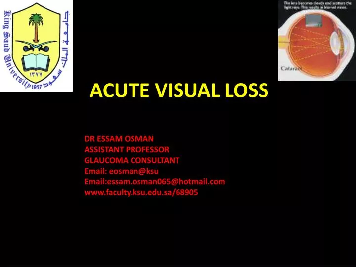
ACUTE VISUAL LOSS. DR ESSAM OSMAN ASSISTANT PROFESSOR GLAUCOMA CONSULTANT Email: eosman@ksu Email:essam.osman065@hotmail.com www.faculty.ksu.edu.sa/68905. ACUTE VISUAL LOSS.

E N D
ACUTE VISUAL LOSS DR ESSAM OSMAN ASSISTANT PROFESSOR GLAUCOMA CONSULTANT Email: eosman@ksu Email:essam.osman065@hotmail.com www.faculty.ksu.edu.sa/68905
ACUTE VISUAL LOSS Sudden onset of blindness is a disaster for most people and you should be able to evaluate such a patient and be able to recognize situations requiring urgent action.
ACUTE VISUAL LOSS 1. Media opacities 2. Retinal disease 3. Optic nerve disease 4. Visual pathway disorders 5. Functional disorders 6. Acute discovery of chronic visual loss
ACUTE VISUAL LOSS Acute glaucoma Central retinal artery occlusion Central retinal vein occlusion Retinal detachment Optic neuritis
ACUTE VISUAL LOSS History: The history questions to be asked of a patient of sudden visual loss include: 1. Is the visual loss transient or persistent? 2. Is the visual loss monocular or binocular? 3. Did the visual loss occur suddenly or it developed over hours, days or weeks? 4. What is the patient’s age and general medical condition? 5.Did the patient have normal vision in the past and when was vision last tested last, as quite a number of people will realize loss of vision from one eye when they cover the good eye.
ACUTE VISUAL LOSS following tests: - Visual acuity testing - Confrontation visual fields - Pupillary reactions - Ophthalmoscopy - External examination of the eye with a pen light - Tonometry to measure the intraocular pressure
Media opacities Corneal edema The cornea appears like a ground glass rather than its normal clear appearance. The most common cause of corneal edema is increased intraocular pressure and occurs typically in angle closure glaucoma. Any acute infection of the cornea by a corneal ulcer may mimic corneal edema.
Media opacities Hyphema Hyphemais blood in the anterior chamber Thehyphemais a direct consequence of blunt trauma to a normal eye. However, it can occur with tumors, diabetes,intraocularsurgery and chronic inflammationwhich all cause neovascularization.
Media opacities Vitreous hemorrhage Any bleeding into vitreous will also reduce the visual acuity. after trauma, seen in diabetics or after a retinal vein occlusion and it may also accompany subarachnoid hemorrhage. If you cannot appreciate a red reflex with an ophthalmoscope and the lens appears clear, you should suspect a vitreous hemorrhage. The diagnosis is confirmed with slit lamp examination through a dilated pupil. B scan is important.
Retinal diseases Retinal detachment an extensive retinal detachment involving the macular area would produce acute visual loss and this patient will complain of flashing lights followed by a large number of floaters and then a shade or blind covering the visual field. An afferent pupillary defect is usually present. The diagnosis is confirmed by ophthalmoscopy through a dilated pupil, and retina appears elevated with folds and the choroidal background is indistinct.
Retinal vascular occlusion Central Retinal artery occlusion – A sudden, painless and often complete visual loss may indicate central retinal artery occlusion. Several hours after a central retinal artery occlusion, the inner layer of the retina becomes opalescent. A cherry red spot is seen due to the pallor of the perifoveal retina in contrast to the normal color of the fovea. A chronic cherry red spot is also a feature of the storage diseases such as Tay-Sachs Pick diseas disease and Niemann-Pick disease.
Retinal vascular occlusion Retinal vein occlusion ophthalmoscopes picture of disc swelling, venous engorgement, cotton wool spots and diffuse retinal hemorrhages like blood and thunder. Loss of vision may be severe. There is no generally accepted acute management. Central retinal vein occlusion is not a true ophthalmic emergency
Retinal vascular occlusion Branch Retinal Artery Occlusion when only a branch of the central retinal artery is occluded, vision is only partially lost. This is more likely to be the result of an emboli and the source of the emboli should be sought. If the visual acuity is affected, attempts should be made to dislodge the emboli by ocular massage.
Optic Nerve Disease Optic Neuritis: Optic Neuritis is inflammation of the optic nerve and is usually associated with multiple sclerosis in a significant number. The visual acuity is markedly reduced and an afferent pupillary defect is present. The optic disc initially appears hyperemic and swollen. The visual acuity usually recovers; however, repeated episodes of optic neuritis may lead to permanent loss ofvision.
Visual Pathway Disorders Homonymous hemianopia - is loss of vision on one side of both visual fields and may result from occlusion of one of the posterior cerebral arteries withinfarctionof the occipital lobe. Other vascular abnormalities occurring in the middle cerebral artery distribution may produce a hemianopia, but usually otherneurologicalsigns are prominent. Any patient with a hemianopia needs at CT or MRI to localize and identify the cause.
Cortical Blindness Cortical Blindness: A rare extensive bilateral damage to the cerebral visual pathways results in complete loss of Vision. This condition is referred to as cortical, central or cerebral blindness. As the pathways serving the pupillary lights reflex separate from those carrying visual information at the level of the optic tracts, a patient who is cortically blind has normal pupillary reactions. Thusa patient with normal fundus examination along with normal pupillaryreactions,mostlikely has cortical blindness..
Functional Disorders A functional disorder is used in preference to hysterical or malingering to describe visual loss without organic basis. A patient may report complete blindness in one eye and normal vision in the other eye, and no relative afferent pupillary defect