Download
1 / 38
460 likes | 1.3k Vues
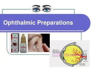
Ophthalmic and Otic Medications. Chapter 18. Basic Anatomy & Physiology. The ocular system is responsible for vision The ocular system is comprised of the eyes and adnexa Globe consists of three layers:

E N D
Ophthalmic and Otic Medications Chapter 18
Basic Anatomy & Physiology • The ocular system is responsible for vision • The ocular system is comprised of the eyes and adnexa • Globe consists of three layers: • Sclera (white of the eye), choroid (middle, vascular coat of the eye), and retina (“the film in the camera”) • Adnexa consists of the surrounding structures: • Orbit, eye muscles, eyelids, eyelashes, conjunctiva, and lacrimal apparatus
Ophthalmic Drugs • Things to consider when using topical ophthalmic drugs • Drug penetration - They must be absorbed into the anterior chamber • Frequency of drug application - They may be administered at different frequencies depending on whether they are in ointment or solution form • Ease of application - They must be relatively easy to administer so that client compliance occurs
Diagnostic Ophthalmic Drugs • Topical anesthetics such as proparacine and tetracaine are used to help perform comprehensive eye exams or to remove foreign material from the eye. • Corneal anesthesia is accomplished in about one minute and lasts for about ten minutes. • Store open bottles in refrigerator. • Discard discolored solutions. • Do not apply before performing STT!!!!
Diagnostic Ophthalmic Drugs • Fluorescein sodium stain is applied to the cornea (using sterile saline) to assess any corneal defects (the stain is orange until it adheres to a corneal defect, where it appears green) • Stain is fat soluble and therefore unable to penetrate or adhere to intact cornea (can only penetrate damaged tissues) • Stain should be washed from the eye before and after examination is complete. • Use a Wood’s Lamp to examine eye for abrasions.
Miotics • Cholinergic drugs that constrict the pupil • Used to treat open-angle glaucoma because they decrease intraocular pressure. • An example is pilocarpine (Piloptic®)
Mitotics vs. Mydriatics • Mitoticsare cholinergic or sympatholytic drugs. • Mydriaticsare sympathomimetic or anticholinergic drugs.
Mydriatics and Cycloplegics • Mydriaticsdilate the pupil and are used to aid in eye exams, to relieve inflammation associated with uveitis (inflammation of the iris, ciliary body, and choroid) and keratitis (inflammation of the cornea), to break up or prevent adhesions between the iris and the lens, and to prepare an animal for ocular surgery • Cycloplegics paralyze the ciliary muscles and minimize pain
Atropine • Anticholinergic drug used for treatment of acute inflammation of anterior uvea and aid in exam of retina • Mydriasisand cycloplegia • Side effects = salivation • Contraindications: glaucoma (increases intraocular pressure); KCS (decreases tear production) • Solution or ointment
Homatropine • Same uses, side effects, and contraindications as atropine. • Faster onset and shorter duration of action than atropine • IsoptoHomatropine®
Phenylephrine • Sympathomimetic drug used to evaluate eye diseases such as uveitis and Horner’s syndrome • May be used prior to conjunctival surgery to decrease hemorrhage • Mydriasis/no cycoplegia • Produces vasoconstriction, ocular discomfort, tearing, and rebound miosis • Mydfrin®
Horner’s Syndrome • Enophthalmos (backward displacement of eyeball into the orbit) • Ptosis of the upper eyelid • Slight elevation of the lower lid • Constriction of pupil • Narrowing of palpebral fissure
Tropicamide • Used for fundic examination • Rapid acting mydriaticwith slight cycloplegic effect. • More rapid onset and shorter duration of action than atropine. • Side effects: local discomfort and salivation • Contraindicated in animals with glaucoma or KCS
Epinephrine • Sympathomimetic drug that reduces intraocular pressure and produces mydriasis. • Used to prevent glaucoma in the unaffected eye • May cause ocular discomfort.
Glaucoma • Glaucoma is a group of diseases that increase intraocular pressure (drugs used to treat glaucoma decrease intraocular pressure) • Primary glaucoma: caused by an acquired structural defect within the eye • Secondary glaucoma: a consequence of another ocular disease or trauma • Congenital glaucoma: resulting from a genetic defect • If left untreated, glaucoma can result in blindness.
Narrow vs Open-Angle Glaucoma • Aqueous humor is constantly produced by ciliary process behind iris • Production is controlled by carbonic anhydrase • Once aqueous humor enters eye, it passes from posterior chamber, through pupil, into anterior chamber, and is then drained through trabecular meshwork. • When intraocular pressure increases, outflow mechanism for aqueous humor is blocked
Narrow vs. Open-Angle Glaucoma (cont’d) • If iris occludes trabecular meshwork, normal outflow of humor is prevented, and animal is said to have narrow-angle glaucoma. • If there is no change in chamber angle of eye, but aqueous humor outflow is impeded because of degenerative changes, animal is said to have open-angle glaucoma.
Drugs Used to Treat Glaucoma • Miotics: covered previously • Carbonic anhydrase inhibitors interfere with the production of carbonic acid, leading to a decrease of aqueous humor production • Examples include acetazolamide, dichlorphenamide, and methazolamide • Beta-adrenergic blockers decrease production of aqueous humor. Systemic side effects (bradycardia, hypotension, bronchospasms). Used with primary glaucoma to prevent development of disease in both eyes. May cause blurred vision. • Examples include timololmaleate and betaxolol hydrochloride
Drugs Used to Treat Glaucoma • Osmotic diuretics: used prior to surgery or as an emergency treatment of glaucoma. • Given IV to decrease vitreous humor volume and rapidly decrease intraocular pressure • Side effects: electrolyte imbalances, cardiovascular problems, vomiting • Examples include mannitol and glycerin
Drugs used to treat KCS • KCS is a disease in which tear production is decreased, resulting in mucopurulent conjunctivitis and corneal scarring/ulceration • Examples of drugs used to treat KCS: • Artificial tears • Antibiotic-steroid preparations • Lacrimogenics (increase tear production) such as pilocarpine • Immunomodulators (interfere with interleukin production by T-lymphocytes) such as cyclosporine
Other Ophthalmic Drugs • Other ophthalmic drugs used to treat ocular diseases include: • Antibiotics • Antifungals • Antivirals • Corticosteroids • NSAIDs • Tear supplements • See Table 18-1 in your textbook for a list of anti-infectives, anti-inflammatories, and tear supplements used in veterinary medicine
Basic Anatomy & Physiology • The ear is the sensory organ that allows hearing and maintains balance • The ear is comprised of three parts: • Outer: pinna and external auditory canal • Middle: tympanic membrane, auditory ossicles, eustachian tube, oval window, and round window • Inner: vestibule, cochlea, and semicircular canals • Otitis interna is an inner ear infection • Side effects include head tilt toward the infected side, ataxia, nausea, and vomiting
Otic Medications • Many drug combinations are used in veterinary medicine to treat ear disease, including: • Antibiotics • Antiparasitics • Antifungals • Corticosteroids (in combination with anti-infectives) • Otic drying agents • Otic cleansing agents • Oticdewaxingagents (cerumen = earwax) • Refer to Table 18-2 in your textbook for a complete list of otic drugs
Epi-Otic & Cerulytic Propylene glycol & salicylic acid Ceruminolytic (propylene glycol, benzyl alcohol)