Download
1 / 6
210 likes | 716 Vues
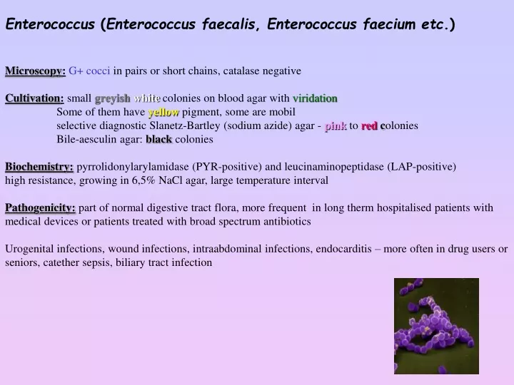
Enterococcus ( Enterococcus faecalis , Enterococcus faecium etc .) M icroscopy : G+ cocci in pairs or short chains, c atal ase negativ e C ultiva tion : small greyish white c oloni es on blood agar with virida tion Some of them have yellow pigment , some are mobil

E N D
Enterococcus (Enterococcus faecalis, Enterococcus faecium etc.) Microscopy:G+ cocciin pairsor short chains, catalase negative Cultivation:smallgreyish whitecolonieson blood agar withviridation Some of them have yellow pigment, some are mobil selective diagnosticSlanetz-Bartley (sodium azide)agar - pinktoredcolonies Bile-aesculin agar: blackcolonies Biochemistry: pyrrolidonylarylamidase (PYR-positive) andleucinaminopeptidase (LAP-positive) high resistance, growing in 6,5% NaCl agar, large temperature interval Pathogenicity:part of normal digestive tract flora, more frequent inlong therm hospitalised patients with medical devicesorpatients treatedwith broad spectrum antibiotics Urogenital infections, wound infections,intraabdominalinfections, endocarditis – more often in drug usersorseniors, catether sepsis, biliary tract infection
Factorsof virulence: gelatinase,feromonsubstance,colonizationfactors,bacteriocins - inhibitionof other bacteria VanA, B, C gens causes rezistenceto vancomycin (Cis gen of primary resistence, VanA/Bof secondary resistance, transferable through plasmides) Treatment:primary resistantto cefalosporines Lihturinary tract infection: ampicillin, ampicillin with–lactamase inhibitors, nitrofurantoin, possible glycopeptides. Wound infections, sepsis and endocarditis: combinationof aminoglykoside+ penicillin/ampicillin or glycopeptides (vancomycin, teicoplanin) VRE (vancomycin resistant enterococci) – linezolid, quinupristin/dalfopristin Laboratory dg.: microscopy, cultivation onBA, on Slanetz-Bartley medium Latex agglutination – differentiation from streptococci, from other bacteriathrough PYR test and LAP Phenotypic test (productionof yellow pigment, moovement) Biochemistry: fermentation of arabinosis and pyruvate: E. faeciumE. faecalis arabinosis fermentation – change of the indicators colourwithout fermentation pyruvate negative pyruvatefermentation resistentto ampicilin susceptibleto ampicillin EN-coccus test
Hysteria? G+ rods Listeria monocytogenes Morphology:microscopy: G+rods, catalase positive Cultivation: chromogennousmedia, growth incold, on BA formgreycolonieswith haemolysis – looks like enterococci, streptococcior difteroids Pathogenicity: wound infection, new-born babies infection (meningitisor sepsis) Virulence factors: lysteriolysin, internalins (intracellular alive) Treatment:fluoroquinolons Laboratory dg.: microscopy, cultivationon chr. medium/ BA andbile-aesculin medium,catalase detection, BBL test Don´t eat Listeria
Corynebacterium difteriae Microscopy:G+rodswith metachromatic granules,club-shapedlooking likechineese signs, catalasa positive Cultivation:does not grow on MH, butonBA, on telur media (Clauberg) Pathogenicity:strains producing toxin (microb attacked by fag) causesdiphteriawith pablanes(couldn´t take offwithoutbleeding), man suffocate, arise of myocarditisetc. Non-toxic strainscausesskininflammations. Factorsof virulence: diphteric toxin Therapy:vaccination, antidiphteric globulin (deserters!), PNC, tracheostomy, cortikoids Laboratory dg.: microscopy, staining of specific parts - granules (Lebranc), Clauberg medium - metalshinycolonieswithblue zone around colonies, Lofler medium, detection of toxinsthrough Elek test, PCR, demonstrationonguinea-pig. OtherCorynebacteria (C. jejkeium etc.) Microscopy:G+ rods with metachromat. granules, club-shapedformlooks likechineesesigns, arranged in palisades, catalase positive Cultivation:any growth on MH, butBA Pathogenicity:wound infection, sepsis, urinary tract infections Factors of virulence: haemolysins Treatment:vancomycin, teicoplanin, rifampicin, if posssible - PNC Laboratory dg.: microscopy, cultivationon BA, biochemistry…
Rod Bacillus B. antracis Microscopy:G+rodslooks like bamboo stick, spors (central terminated) – only in air Cultivation:onBA – large, flat, spreading through the agar surface - caput medusae, ahaemolytical Pathogenicity and pathogenesis: contact withill person, dead animalsortheir productes (skin), spors invade into organism,germinate and produce toxin. Via entrance is disease devided into3 forms. 1. skin - pustula maligna 2. pulminal – after inhalationarises hemoragic necrosisof nodeswith mediastinitisends as septicshock 3. intestinal – via contaminatedfood – causesbloodydiarrhea, high temperatureetc. !! sporsare easy to diffuse, that´s why it is discussed asa biologicalwarfare!! Virulence:toxin (3components) Therapy: PNC, ciprofloxacin, doxycyklin, chloramphenicol Prevention:veterinary control of animal, vaccination of animal or people Laboratory diagnosis:microscopy, cultivationon BA Antigen detection - Ascoli termoprecipitation reaction, animal demonstration !! Can do only laboratory withbiosafety level III.
B. cereus Microscopy:G+rods, central terminated spores Cultivation:onBA flatcolonieswith β haemolysis, PEMBA-bluecolonies Pathogenicity: componentof gastrointesinal flora, contamination of food, causingdiarrhea, vomitting. Diarrhea is caused by thermolabil enterotoxin (source:sauce), vomittingis caused by thermostabil toxin (source: rice). Also causeseye + wound infection Factorsof virulence: enterotoxins Treatment: rehydratation + linkosamids. Prevention: goodfood preparation Eye infection: lincosamids + aminoglycosides Laboratory dg.: microscopy, cultivationonBA/PEMBA, detection of granules toxin detection via ELISA method or latex agglutination


![Trivedi Effect - Impact of an external energy on Enterococcus faecalis [ATCC – 51299] in relation to antibiotic suscepti](https://cdn4.slideserve.com/7685708/5-14-2015-dt.jpg)