Download
1 / 171
1.71k likes | 1.8k Vues
Structural Classification of the Nervous System. Central nervous system (CNS) Organs Brain Spinal cord Function Integration; command center Interpret incoming sensory information Issues outgoing instructions. Structural Classification of the Nervous System.

E N D
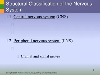
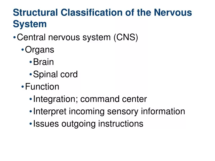
Structural Classification of the Nervous System Central nervous system (CNS) Organs Brain Spinal cord Function Integration; command center Interpret incoming sensory information Issues outgoing instructions
Structural Classification of the Nervous System Peripheral nervous system (PNS) Somatic Nervous System Nerves extending from the brain and spinal cord Spinal nerves—carry impulses to and from the spinal cord(31 pairs) Cranial nerves—carry impulses to and from the brain(12 pairs)) Functions Serve as communication between the brain and spinal cord, and glands or muscles
Functional Classification of the Peripheral Nervous System Autonomic nervous system = involuntary Automatically controls smooth and cardiac muscles and glands Further divided into the sympathetic and parasympathetic nervous systems
Nervous Tissue: Support Cells&neurons Supporting cells in the CNS are grouped together as “neuroglia” General functions Support Insulate Protect neurons
Nervous Tissue: Support Cells 1.Astrocytes Abundant, star-shaped cells Surround neurons Form barrier between capillaries and neurons, BBB,touching the capillaries prevents escape of toxins to brain tissues Control the chemical environment of the brain
Capillary Neuron Astrocyte (a) Astrocytes are the most abundant and versatile neuroglia. Figure 7.3a
Nervous Tissue: Support Cells 2.Microglia Spiderlike phagocytes the main immune cells of the nervous system
Neuron Microglial cell (b) Microglial cells are phagocytes that defend CNS cells. Figure 7.3b
Nervous Tissue: Support Cells 3.Ependymal cells Line cavities of the brain and spinal cord Have cilia assisting in circulation of cerebrospinal fluid
Fluid-filled cavity Ependymal cells Brain or spinal cord tissue (c) Ependymal cells line cerebrospinal fluid-filled cavities. Figure 7.3c
Nervous Tissue: Support Cells 4.Oligodendrocytes Wrap around nerve fibers in the central nervous system Produce myelin sheaths in CNS
Myelin sheath Process of oligodendrocyte Nerve fibers (d) Oligodendrocytes have processes that form myelin sheaths around CNS nerve fibers. Figure 7.3d
Nervous Tissue: Support Cells 5.Schwann cells Form myelin sheath in the peripheral nervous system
Satellite cells Cell body of neuron Schwann cells (forming myelin sheath) Nerve fiber (e) Satellite cells and Schwann cells (which form myelin) surround neurons in the PNS. Figure 7.3e
Nervous Tissue: Neurons Neurons specialized to transmit messages Major regions of neurons Cell body—nucleus and metabolic center of the cell Processes—fibers that extend from the cell body (denderites)
Nervous Tissue: Neurons Cell body has: Nissl bodies(granules) Specialized rough endoplasmic reticulum Neurofibrils cytoskeleton Maintains cell shape Nucleus with large nucleolus
Cell body Dendrite Mitochondrion Nissl substance Axon hillock Axon Collateral branch Neurofibrils Nucleus One Schwann cell Node of Ranvier Axon terminal Schwann cells, forming the myelin sheath on axon (a) Figure 7.4a
Neuron cell body Dendrite (b) Figure 7.4b
Nervous Tissue: Neurons Processes outside the cell body Dendrites—conduct impulses toward the cell body Neurons may have hundreds of dendrites Axons—conduct impulses away from the cell body Neurons have only one axon arising from the cell body at the axon hillock
Cell body Dendrite Mitochondrion Nissl substance Axon hillock Axon Collateral branch Neurofibrils Nucleus One Schwann cell Node of Ranvier Axon terminal Schwann cells, forming the myelin sheath on axon (a) Figure 7.4a
Nervous Tissue: Neurons Axons End in axon terminals Axon terminals contain vesicles with neurotransmitters Axon terminals are separated from the next neuron by a gap Synaptic cleft—gap between adjacent neurons Synapse—junction between nerves
Nervous Tissue: Neurons Myelin sheath—whitish, fatty material covering axons Schwann cells—produce myelin sheaths in jelly roll-like fashion around axons (PNS) Nodes of Ranvier—gaps in myelin sheath along the axon Oligodendrocytes—produce myelin sheaths around axons of the CNS
Schwann cell cytoplasm Schwann cell plasma membrane Axon Schwann cell nucleus (a) (b) Neurilemma Myelin sheath (c) Figure 7.5
Neuron Cell Body Location Most neuron cell bodies are found in the central nervous system Nuclei—clusters of cell bodies within the white matter of the central nervous system Ganglia—collections of cell bodies outside the central nervous system
Neuron Cell Body Location Tracts—bundles of nerve fibers in the CNS Nerves—bundles of nerve fibers in the PNS White matter—collections of myelinated fibers (tracts) Gray matter—collections of mostly unmyelinated fibers and cell bodies
Functional Classification of Neurons Sensory (afferent) neurons Carry impulses from the sensory receptors to the CNS Motor (efferent) neurons Carry impulses from the central nervous system to viscera, muscles, or glands
Central process (axon) Sensory neuron Spinal cord (central nervous system) Cell body Ganglion Dendrites Peripheral process (axon) Afferent transmission Interneuron (association neuron) Peripheral nervous system Receptors Efferent transmission Motor neuron To effectors (muscles and glands) Figure 7.6
Functional Classification of Neurons Interneurons (association neurons) Found in the central nervous system Connect sensory and motor neurons
Central process (axon) Sensory neuron Spinal cord (central nervous system) Cell body Ganglion Dendrites Peripheral process (axon) Afferent transmission Interneuron (association neuron) Peripheral nervous system Receptors Efferent transmission Motor neuron To effectors (muscles and glands) Figure 7.6
Structural Classification of Neurons Multipolar neurons—many extensions from the cell body All motor and interneurons are multipolar Most common structure
Cell body Axon Dendrites (a) Multipolar neuron Figure 7.8a
Structural Classification of Neurons Bipolar neurons—one axon and one dendrite Located in special sense organs such as nose and eye Rare in adults
Cell body Axon Dendrite (b) Bipolar neuron Figure 7.8b
Structural Classification of Neurons Unipolar neurons—have a short single process leaving the cell body Sensory neurons found in PNS ganglia
Dendrites Cell body Short single process Axon Central process Peripheral process (c) Unipolar neuron Figure 7.8c
Functional Properties of Neurons Irritability Ability to respond to stimuli Conductivity Ability to transmit an impulse
Nerve Impulses Resting neuron The plasma membrane at rest is polarized Fewer positive ions are inside the cell than outside the cell
[Na+] + + + + + + + 1 + Resting membrane is polarized. In the resting state, the external face of the membrane is slightly positive; its internal face is slightly negative. The chief extracellular ion is sodium (Na+), whereas the chief intracellular ion is potassium (K+). The membrane is relatively impermeable to both ions. – – – – – – – – [K+] – – – – – – – + + + + + + + Figure 7.9, step 1
Nerve Impulses Depolarization A stimulus depolarizes the neuron’s membrane The membrane is now permeable to sodium as sodium channels open A depolarized membrane allows sodium (Na+) to flow inside the membrane
Na+ + + + + + + + 2 Stimulus initiates local depolarization. A stimulus changes the permeability of a local "patch" of the membrane, and sodium ions diffuse rapidly into the cell. This changes the polarity of the membrane (the inside becomes more positive; the outside becomes more negative) at that site. + – – – – – – – – + – – – – – – – + + + + + + + Figure 7.9, step 2
Nerve Impulses Action potential The movement of ions initiates an action potential in the neuron due to a stimulus A graded potential (localized depolarization) exists where the inside of the membrane is more positive and the outside is less positive
Na+ + + – + + – + 3 + Depolarization and generation of an action potential. If the stimulus is strong enough, depolarization causes membrane polarity to be completely reversed and an action potential is initiated. – – – – – + + – + – – – – – + + – + + – + + + Figure 7.9, step 3
Nerve Impulses Propagation of the action potential If enough sodium enters the cell, the action potential (nerve impulse) starts and is propagated over the entire axon Impulses travel faster when fibers have a myelin sheath
– – – + – – + + 4 Propagation of the action potential. Depolarization of the first membrane patch causes permeability changes in the adjacent membrane, and the events described in step are repeated. Thus, the action potential propagates rapidly along the entire length of the membrane. + – + – + – + + 2 – + – + + – + – – – + – + + Figure 7.9, step 4
Nerve Impulses Repolarization Potassium ions rush out of the neuron after sodium ions rush in, which repolarizes the membrane Repolarization involves restoring the inside of the membrane to a negative charge and the outer surface to a positive charge