Download
1 / 61
5.28k likes | 16.83k Vues
CIRRHOSIS OF LIVER . Dr.Vemuri Chaitanya. Cirrhosis. Chronic generalized liver disease A condition that is defined histopathologically and has a variety of clinical manifestations and complications, some of which can be life threatening.

E N D
CIRRHOSIS OF LIVER Dr.Vemuri Chaitanya
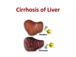
Cirrhosis • Chronic generalized liver disease • A condition that is defined histopathologically and has a variety of clinical manifestations and complications, some of which can be life threatening. • Pathologic features : development of fibrosis to the point that there is architectural distortion with formation of regenerative nodules ( micronodular / macronodular ) • This results in decrease in hepatocellular mass, thus function .
Epidemiology • 40% cases asymptomatic • It is the 12th leading cause of death in United States. • Approximately 30,000 to 50,000 deaths per year • Additional 10,000 deaths due to liver cancer secondary to cirrhosis
This end stage of CLD is characterised by : • Bridging Fibrous Septa • Parenchymal nodules • Disruption of the architecture of the entire liver
pathogenesis • Hepatocellular death • Regeneration • Progressive fibrosis • The induction of fibrosis occurs with activation of hepatic stellate cells, resulting in formation of increased amounts of collagen & other components of extracellular matrix. • Stimuli : 1.Chr.inflammation – cytokines like TNF, Lymphotoxin, IL-1 2.Cytokine production by injured Kupffer cells, endothelial cells, hepatocytes, bile duct epithelial cells 3.Disruption of ECM 4.Direct stimulation of stellate cells by toxins
Etiology • Alcoholism • Chronic Viral Hepatitis – Hepatitis B Hepatitis C • Autoimmune Hepatitis • Nonalcoholic steatohepatitis • Biliary Cirrhosis – Primary biliary cirrhosis Primary sclerosing cholangitis Autoimmune cholangiopathy
Etiology • Cardiac Cirrhosis • Budd Chiari Syndrome • Inherited metabolic liver disease : Hemochromatosis Wilson’s Disease Alpha 1 Antitrypsin deficiency Cystic Fibrosis • Cryptogenic Cirrhosis • Others : Galactosemia , Tyrosinemia, Drug induced : alpha methyldopa Syphilis
Clinical Features • Asymptomatic for long periods. • Onset of symptoms – insidious , less often abrupt. • Non specific symptoms – vague right upper quadrant pain, fever, nausea, vomiting,diarrhea,anorexia & malaise. • Or they may present with more specific complication of CLD – ascites,upper GI bleed etc…
Signs • Loss of hair ( alopecia ) • Icterus • Pallor • KF Ring • Parotid enlargement • Fetor hepaticus • Loss of axillary & pubic hair • Spider nevi • Gynecomastia • Atrophy of breasts in females • Wasting of muscles
Signs • Glossitis, cheilitis • Palmar erythema • Clubbing • Leuconychia • Dupuytren’s contracture • Ascites • In 70 % cases liver is enlarged, firm if not hard and nodular • Splenomegaly • Caput medusae
Signs • Bleeding tendencies : deficiency of clotting factors – check PT /INR • Fever • Hyperpigmentation • Hyperdynamic circulatory state • Edema • Hernia • Testicular atrophy • Delirium • Constructional apraxia • Flapping tremors • Inversion of sleep rhythm
Alcoholic Cirrhosis • Accurate history regarding amount & duration of alcohol consumption is required. • Lab tests : • Completely normal in early compensated alcoholic cirrhosis • Hb: Anemia + ( chr.GI loss, nutritional def, hypersplenism ) • Platelet count : reduced early in disease, portal htn with hypersplenism
Alcoholic Cirrhosis • S.Bilirubin – normal / elevated • PT – often prolonged • S. Transaminases – elevated • AST / ALT = > 2/1 • Liver biopsy • Treatment : • Abstinence is the cornerstone of therapy. • Treatment of any complications • Glucocorticoids – if DF > 32 • Oral Pentoxiphylline
Cirrhosis d/t Chr.Hepatitis B & C • Of patients exposed to HCV, approximately 80% develop Chronic hepatitis and of those, about 20 –30 % will develop cirrhosis over 20-30 yrs. • Here , liver is small & shrunken with a characteristic features of mixed micro and macro nodular cirrhosis seen on biopsy. • Of patients exposed to HBV, about 5 % develop chronic hepatitis & about 20% of those patients go on to develop cirrhosis. • Liver is small & shrunken and has mixed micro & macronodular cirrhotic pattern. • Invg : Routine investigations + HCV RNA, HBsAg, anti – HBs, HBeAg, anti – HBe, HEV DNA.
Cirrhosis d/t Chr.Hepatitis B & C • Specific Treatment : • HBV : Lamivudine, Adefovir, Entecavir, Tenofovir • HCV: Pegylated Interferon, Ribavirin
Primary Biliary Cirrhosis • Female preponderance • Median age of around 50 yrs • Etiology : unknown • Portal inflammation & necrosis of cholangiocytes in small and medium sized bile ducts. • Antimitochondrial antibodies in 90% of pts • Pathology : earliest lesion- Chronic Nonsuppurative Destructive Cholangitis
Primary Biliary Cirrhosis • Fatigue • Pruritis • On ex: hepatomegaly splenomegaly ascites edema • Unique to PBC : Hyperpigmentation,Xanthelasma,Xanthomata
Primary Biliary Cirrhosis LAB : • Elevated GGT, ALP with mild elevations of AST & ALT • Hyperbilirubinemia • Thrombocytopenia, leukopenia, anemia TREATMENT : • UDCA @ 13 – 15 mg/Kg per day • Liver Transplantation • Cholestyramine • Bisphosphonates – osteopenia/osteoporosis
Primary Sclerosing Cholangitis • Etiology : unknown • Diffuse inflammation & fibrosis of entire biliary tree – chronic cholestasis – obliteration of intra & extrahepatic biliary tree – biliary cirrhosis – portal htn – liver failure • Cli fea : fatigue, pruritis, steatorrhea, fat sol vitamin deficiencies • Lab: 2 fold rise in ALP, elevated aminotransferases, p-ANCA ( 65%) • Diagnosis : MRCP , Cholangiogram • Treatment : No proven treatment. High dose 20 mg/kg/day UDCA. Endoscopic dilatation of dominant strictures Liver Transplantation
Cardiac Cirrhosis • Pts with long standing right sided CHF may develop chronic liver disease & cardiac Cirrhosis • Cli fea : symptoms of Rt.Heart.Failure + Hepatomegaly • Lab : ALP raised, AST > ALT – normal / raised • Diagnosis : cardiac case with elevated ALP & enlarged liver
Cirrhosis – other causes • Hemochromatosis • Wilson’s Disease • Alpha1 Antitrypsin Deficiency • Cystic Fibrosis
Investigations • Complete Hemogram • Peripheral Smear • Platelet Count • PT INR • LFT – S. Bilirubin, S. Albumin, S. Globulin, SGPT, SGOT, ALP • Hepatitis Profile • Alpha Fetoprotein
Investigations • Blood sugar • Urea, Creatinine • Sodium, Potassium • Ascitic fluid examination • X-Ray chest • USG / CT Abdomen • Confirmation by Liver Biopsy
Complications of Cirrhosis • Portal HTN – Gastroesophageal Varices Portal hypertensive Gastropathy Splenomegaly, Hypersplenism Ascites – SBP • Hepatorenal Syndrome – Type 1 & 2 • Hepatic Encephalopathy • Hepatopulmonary Syndrome • Portopulmonary Hypertension • Malnutrition
Complications of Cirrhosis • Coagulopathy : Factor deficiency Fibrinolysis Thrombocytopenia • Bone Disease : Osteopenia/Osteoporosis/ Osteomalacia • Haematological abn : Anaemia Hemolysis Thrombocytopenia Neutropenia
Portal Hypertension • Prehepatic :Portal Vein thrombosis Splenic Veinf Thrombosis Massive Splenomegaly • Hepatic : Presinusoidal – Schistosomiasis Cong.hepatic fibrosis Sinusoidal – Cirrhosis Alcoholic hepatitis Postsinusoidal – Veno-occlusive Disease
Posthepatic : Budd Chiari syndrome Inferior vena caval webs Cardiac Causes – Restrictive Cardiomyopathy Constrictive Pericarditis Severe CHF
Portal Hypertension • Elevation of hepatic venous pressure gradient to > 5mm Hg. • It is caused by combination of 2 simultaneously occuring hemodynamic processes : • Increased intrahepatic resistance to passage of blood flow through liver • Increased splanchnic blood flow secondary to vasodilation.
Portal Hypertension • Portal HTN directly responsible for 2 complications – variceal haemorrhage and ascites • Also hypersplenism, • congestive gastropathy, • renal failure and • hepatic encephaopathy
CLINICAL FEATURES • Splenomegaly – Hypersplenism – Thrombocytopenia, Neutropenia, Anemia • Dilated Abdominal Veins, Caput Medusae, Ascitis. • Oesophageal varices
Variceal Bleed • Approx 5 – 15 % of cirrhotics per year develop varices and it is estimated that majority of patients with cirrhosis will develop varices over their lifetime • 1/3rd of patients with varices develop bleeding. • Factors predicting variceal bleed : • Severity of cirrhosis ( Child’s Class ) • Ht of wedged hepatic vein pressure • Size and location of varix • Endoscopic stigmata : red wale sign, hematocystic spots, diffuse erythema, bluish color, cherry red spot & white nipple spot • Tense ascites
Variceal Bleed • Diagnosis : identified by endoscopy • Pt with a gradient of >12 mm Hg – are at a greater risk for variceal bleed. • Precipitating Factors – Alcohol, Aspirin, Analgesics (NSAIDs), Adrenal Corticosteroids • Assessment – Drop in systolic BP > 10 mmHg, rise in pulse > 15 beats / minute on sitting up – 10 to 20% • Supine Hypotension - > 20% • Systolic BP < 100 mmHg / Baseline Tachycardia > 25%
RESUSCITATION • Stabilize BP – 2 large bore IV line – Isotonic saline / Ringer Lactate / fresh blood / packed RBC transfusion – Maintain ½ hour pulse, BP, respiration chart (In emergency situation – O-ve blood) • MEASURE URINE OUTPUT • Correction of coagulopathy – FFP, parenteral Vit K 10 mg • Platelet transfusion – if count < 50,000 • Airway protection – endotracheal intubation to prevent aspiration
RESUSCITATION • Nasal Gastric Aspiration • OCTREOTIDE Infusion 50 to 100 µgm bolus 25 to 50 µgm / hour infusion • VASOPRESSIN 0.3 unit / minute IV – gradually increased to 0.9 units/minute – Side-effects : Myocardial ischemia, infarction, arrhythmia, cardiac arrest, mesenteric ischemia ( now not preferred )
Resuscitation • Endoscopic Therapy : Variceal band ligation Variceal sclerotherapy • Balloon tamponade ( Sengstaken-Blakemore tube or Minnesota tube ) – in pts who cannot get endoscopic therapy or those who need stabilization prior to endoscopic therapy • TIPS – When esophageal varices extend into proximal stomach In Pts eho fail endoscopic / medical treatment and also poor subjects for surgery.
prophylaxis • Beta blockers – propranolol – resting heart rate to be reduced by 25 %. • Repeated variceal band ligation until varices are obliterated.
Splenomegaly & Hypersplenism • Congestive splenomegaly is common in pts with portal htn. • Clinical features include enlarged spleen, thrombocytopenia, leukopenia • Some – significant left sided/ left upper quadrant abdominal pain • No specific treatment • Splenectomy • Hypersplenism with development of thrombocytopenia – first indicator of portal hypertension
Ascites • Accumulation of fluid within the peritoneal cavity • M.C cause : cirrhosis with portal hypertension • Clinical features : increase in abdominal girth peripheral edema dyspnea – if massive bulging flanks shifting dullness fluid thrill • Hepatic hydrothorax – more common on rt.side implicates rent in diaphragm with free flow of ascitic fluid into thoracic cavity
Ascites • Diagnostic paracentesis • SAAG : • >1.1g/dL – portal hypertension • <1.1g/Dl – neoplasm,Tb, pancreatitis, • Ascitic fluid proteins low – high chance of developing SBP • Ascitic fluid – high RBCs – traumatic tap, HCC, ruptured omental varix • Ascitic fluid – PMN >250 /cu.mm - SBP
Ascites - Treatment • Small amounts of ascites – dietary sodium restriction ( <2g/day ) • Moderate : diuretic is essential Spiranolactone 100-200 mg/day OD Furosemide 40-80 mg/day - if peripheral edema + • Pt is compliant but ascitic fluid + , then Spiranolactone 400 -600 mg/day Furosemide 120-160 mg/day • If ascites still + , then it is REFRACTORY ASCITES
Ascites - treatment • Refractory ascites – Large volume paracentesis TIPS Liver Transplantation • Prognosis – pts of cirrhosis with ascites- poor • <50 % of pts survive 2 yrs after the onset of ascites.
Spontaneous Bacterial Peritonitis • Spontaneous infection of ascitic fluid without any intraabdominal source. • Bacterial translocation – gut flora transversing the intestine into mesenteric lymph nodes, leading to bacteremia and seeding of ascitic fluid • MC : E.coli • Others : Step.viridans, Staph.aureus • If > 2 organisms are identified – secondary bacterial peritonitis d/t perforated viscus to be considered • Ascitic fluid PMN > 250/cu.mm
SBP • Pt can present with altered sensorium, elevated WBC, abdominal pain/discomfort • Treatment : cephalosporins • In pts with an episode(s) of SBP and recovered , once –weekly- administration of antibiotic as prophylactic measure
Hepatorenal Syndrome • Functional renal failure without renal pathology • 10% of pts with cirrhosis / advanced liver failure • Diagnosis : presence of large amount of ascites progressive rise in creatinine urinary sodium <10 mEq • Type 1 HRS : progressive impairment of renal function & significant reduction in creatinine clearance within 1- 2 wks . BAD PROGNOSIS • Type 2 HRS : reduction in GFR, with rise in S.Creat BETTER PROGNOSIS
Hepatorenal Syndrome • Seen in refractory ascites • Exclude causes of ARF • Treatment: • Midodrine, an alpha agonist along with Octerotide and IV Albumin • Liver transplantation