Download
1 / 31
310 likes | 493 Vues
Mesoangioblast stem cells ameliorate muscle function in dystrophic dogs. Authors: Source: Nature. 2006 Nov 30 :574-9. Outlines. 1. Background : a. Human Duchenne Muscular Dystrophy ( DMD )

E N D
Mesoangioblast stem cells ameliorate muscle function in dystrophic dogs Authors: Source: Nature. 2006 Nov 30 :574-9.
Outlines 1. Background : a. Human Duchenne Muscular Dystrophy ( DMD ) b. Golden retriever muscular dystrophy model ( GRMD ) c. Mesoangioblast stem cells 2. Experimental design 3. Results 4. Conclusions
Duchenne muscular dystrophy ( DMD ) Genetics: X-linked ( Xp21 ) recessive dystrophin-deficient muscular dystrophy leading to weakened sarcolemma . Clinical features: a. Onset in early childhood b. Progressive muscular weakness ,hard to run and climb stairs c. Gower’s manoeuvre d. Wheelchair needed in most cases by age 12 e. 20 % boys , IQ< 70 f. Epidemiology: 3 x10⁻⁴ at boys birth g. death at cardiac involvement Lab diagnosis: a. Serum creatine kinase: released by damaged muscle fibres b.Electromyography c.Muscle histology : fibre size , fibre necrosis, invasion by macrophages, and replacement by fat and connective tissue. d. immunohistochemistry for dystrophin protein Treatment: a. No effective drugs b. Gene therapy c. Cell therapy Normal DMD dystrophin
Dystrophin protein • rod-shaped structural protein, about 150 nm, 3684 amino acids ,M.W. 427 kDa • Functions: • a. connect the sarcolemmal cytoskeleton to the extra-cellular matrix • b. dissipate muscle contractile force from the intracellular cytoskeleton to the extracellular matrix • Loss of function: • membrane fragility and sarcolemma injury during contraction Muscle membrane Dystrophin glycoprotein complex ( DCG )
Golden retriever muscular dystrophy ( GRMD ) • Human DMD animal model • display clinical signs of human DMD • Great difficulty in walk by 8 months of age and death at 1 year GRMD dog
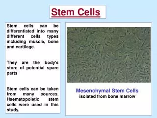
Mesoangioblast stem cells • a class of vessel-associated fetal stem cells • differentiate into most mesoderm cell types when exposed to certain cytokines • more than 50 passages in culture and no tumorigenesis in nude mice Aim: To test the efficacy of stem cell and/or gene therapy in GRMD dogs
Myosin light chain 1 fast promoter rapamycin cyclosporine Experimental design Autologous,gene therapy Heterologous , wild type donor Untreated Lentiviral vector expressing human microdystrophin Muscle-specific creatine kinase promoter Mesoangioblasts GRMD dogs
Morphology Proliferation Wild-type Dystrophic Euploid Karyotype ( 78 chromosomes ) Isolation and characterization of canine mesoangioblasts 15 days postnatal ( P15 )
Lentiviral vector expression of canine mesoangioblasts Mesoangioblasts with lentiviral vector Mesoangioblasts with lentiviral vector + C2C12 mouse myoblasts co-culture Mesoangioblasts Myotubes GFP expression GFP expression
Lentiviral vector expression of canine mesoangioblasts Mesoangioblasts with lentiviral vector + MyoD (Myogenic Determination protein ) transfection Myotubes Myotubes GFP expression MyHC expression ( Myosin Heavy Chain ) Merged
Conclusions • Mesoangioblasts isolated from muscle biopsies were proliferated and differentiated well • in vitro. • Lentiviral vector could be transduced and expressed in mesoangioblasts.
Migration of canine mesoangioblasts into skeletal muscle Mouse mesoangioblasts ( GFP-expressed lentiviral vector ) GRMD mesoangioblasts ( GFP-expressed lentiviral vector ) SCID mice’s femoral artery ( Serve combined immunodeficiency ) 6 hours Isolation of several muscles Real-time PCR analysis for GFP expression
Mouse GRMD dogs Migration of canine mesoangioblasts into skeletal muscle i : Injected leg U : Unjected leg Qd: Quadriceps Gs: Gastrocnemius TA: Tibialis cranialis Lv: Liver Sp: Spleen
Muscle fibres reconstitution of canine mesoangioblasts ※21 days Mouse muscle fibres Mouse muscle fibres Lamin A-C Dystrophin ( Nuclear Envelope Marker ) Laminin DAPI ( Structural protein in membrane )
Conclusions • . Canine mesoangioblasts migrated from the femoral artery to the downstream muscles with • an efficiency similar to that of their wild-type mouse counterparts • Canine mesoangioblasts had the ability to reconstitute muscle fibres
Muscle-specific creatine kinase promoter cyclosporine Autologous V.S. Heterologous cell transplantation Autologous gene therapy Heterologous wild type donor Untreated Lentiviral vector expressing human microdystrophin Mesoangioblasts 3 injections ( 1-month intervals, 5x10⁷ cells ) GRMD dogs
Autologous V.S. Heterologous cell transplantation Autologous Heterologous Morphology Dystrophin Laminin
Conclusions Modified treatment: a. Increase injections to five b. Use stronger myosin light chain 1F promoter
Myocarditis Heterologous cell transplantation Wild-type Mesoangioblasts cyclosporine Rapamycin Rapamycin/ IL-10 GRMD dogs 5 injections 3 injections 3 injections
Heterologouscell transplantation 5 injections & cyclosporine U: unjected leg i : injected leg Morphology Sar: Sartorius Gas: Gastrocnemius TC: Tibialis cranialis BF: Biceps femoralis Dystrophin β- Sarcoglycan Laminin Biceps femoralis
After 13 months 3 injections & Rapamycin 5 injections & cyclosporine Valgus Varus
Conclusions • The clinical motility of GRMD dogs was improved by 5 cell injections . • The Immuno-supression did not show significant differences between cyclosporine and rapamycin. • The heterologous GRMD dogs expressed well-preserved morphology and dystrophin protein . • The expression of β-sarcoglycan indicated reconstitution of the dystrophin-associated complex.
Pneumonia Autologous, modified gene therapy Lentiviral vector expressing human microdystrophin Myosin light chain 1 fast promoter Mesoangioblasts 5 injections GRMD dogs
U: unjected leg i: injected leg Sar: Sartorius Gas: Gastrocnemius TC: Tibialis cranialis BF: Biceps femoralis Autologous, modified gene therapy Before After Morphology β-sarcoglycan Dystrophin Laminin
Conclusions Vampire All three dogs treated with autologous ,genetically corrected cells performed poorly ,even though two of them showed amelioration of morphology and expression of dystrophin protein.
To test less effective results obtained with autologous cells was due to the later onset of the treatment
Azor Azur The efficacy of late transplantation of donor mesoangioblasts Dystrophin Laminin
Conclusions • Even with a later onset of treatment, heterologous cell transplantation seems to produce a • greater amelioration of muscular dystrophy than is produced by autologous dystrophin- • expressing cells .
Normal dog Autologous GRMD dog ( P113 ) Heterologous GRMD dog ( P75 ) Heterologous GRMD dog ( P159 ) Untreated GRMD dog Heterologous GRMD dog ( P75 ) Heterologous GRMD dog ( P159 ) Heterologous GRMD dog ( P159 ) Autologous GRMD dog Force of treated leg Force of untreated leg X 100 % Analysis of enhancement of contraction force in heterologous GRMD dogs A. Tetanic force of skeletal muscles in vivo B. Force of contraction on isolated single muscle fibres in vitro
Immunostaing by dystrophin antibody Specific force Dystrophin expression Heterologous Analysis of enhancement of contraction force in heterologous GRMD dogs Force of contraction on isolated single muscle fibres in vitro
Conclusions • The transplantation of mesoangioblasts into dystrophic cells could obtain an extensive reconstitution of fibres expressing dystrophin ,an improvement in the contraction force and a preservation of walking ability. • Donor wild-type mesoangioblasts seemed to be more efficient than autologous ,genetically corrected cells. • A different onset of treatment should not be crucial. • Mesoangioblasts were a good candidates for future stem cell therapy for Duchenne patients.