Download
1 / 66
660 likes | 841 Vues
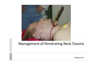
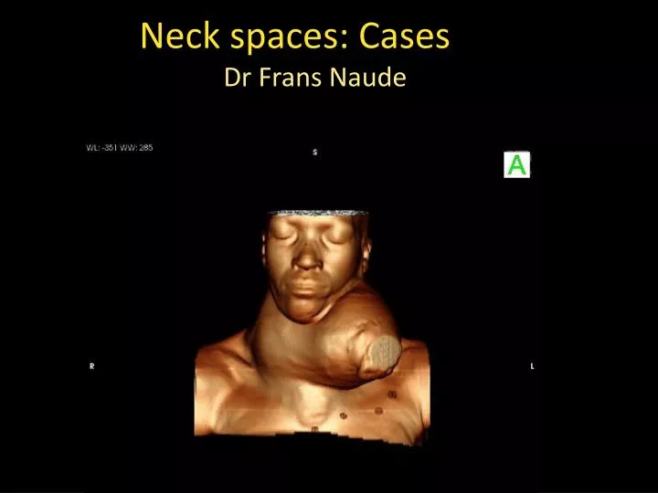
Neck spaces: Cases. Dr Frans Naude. Lesotho patient presented with neck swelling for the last 26 years . 1 avi. Key image 1. Iodine deficient regions Decrease thyroid hormone Increased TSH Goiter Risk of low iodine: increased breast cancer ( Japanese 6/100 000, USA 22/100 000)

E N D
Neck spaces: Cases Dr Frans Naude
Lesotho patient presented with neck swelling for the last 26 years
Iodine deficient regions • Decrease thyroid hormone • Increased TSH • Goiter • Risk of low iodine: increased breast cancer ( Japanese 6/100 000, USA 22/100 000) • Japanese iodine uptake x25 higher
Iodine deficiency is also associated with increased risk for thyroid carcinoma in animal models and humans. • Iodine replacement increase risk ratio from papillary to follicular cancer (Altern Med Rev 2008;13(2):116-127) Ulla Feldt-Rasmussen. Thyroid. May 2001, 11(5): 483-486.
Child with neck mass • UM00421837 • 3.5 year old • Presented with mass in the neck
avi Pt r2 ax
pt r 2 cor Avi
pt r 2 sag l Avi
Expert DDX 5-12 Cystic neck masses in child DIA H&N 5-13
Lymphangioma • Def: Uni/Multiloculated ,non-enhancing cystic neck masses with imperceptible wall that insinuates between vessel and the normal neck structures • Contiguous neck space involvement(trans-spatial) • Synonyms = cystic hygroma/lymphatic malformation
Region • Supra hyoid – submandibular and masticator spaces • Infrahyoid – posterior cervical space • Invaginates into normal structures with minimal mass effect/ multi or uniseptated
CT Findings NECT: Low density, poorly circumscribed cystic neck mass • Fluid –Fluid lesions in multiloculated lesions CECT • No significant enhancement in mass or wall • (complex lesions, veins may cause enhancement)
MRI • T1 w: Primarily hypointense , may be hyperintense due to hemorrhage or protein rich fluid. • Fluid –fluid levels often seen. • T2-w: hyperintense throughout ( best sequence to map lesion) • Trans-spatial extension/poorly marginated. • T1+C: most often no enhancement. If enhancement present ,most likely due to mixed vascular structures
U/S • Confirm diagnosis • Classify type ( macrocysitic/microcytic and mixed) ( microcytic with cyst <1cm ) BiomImagInterv J 2011;7(3): e 18
Take home point • Unilocular cervical lesion : thyroglossal cyst, branchial cleft cysts, thymic cyst • ( lymphangioma = multilocular) • Lymphangioma = trans spatial
Treatment • Surgical • Bleomycinsclerotherapy BiomImagInterv J 2011;7(3): e 18
Cystic neck masses in adult DIA H&N 5-16
Adult with neck mass • 53 yr • Right neck mass
Carotid body tumor • Location : Mass in the center of the carotid bifurcation, splaying the ECA and ICA • Avid enhancing mass DI H&N III 8 :21
pt 2 with cbt Avi
pt 2 w cbtcor Avi
Patient with: • Neuropathy of left cranial nerve 7-12 • Tinnitus
Glomusjugulareparaganglioma • Clinical: Pulsatile tinnitus with vascular retrotympanicmass • Neuropathy : Cranial nerve 9-12 (sometimes 7&8) • Arises from margin of jugular foramen (neural crest cells surrounding the jugular foramen) • Projects supero-laterally into middle ear cavity • Permeativedestructive bony changes on CT • Vertical part of the posterior wall of ICA often involved
DDX: • Jugular foramen schwanoma, meningioma, pseudolesion, metatases
Patient 5 • Stridor, hoarseness, dyspnea • Smoking history • Right neck mass