Download
1 / 16
160 likes | 261 Vues
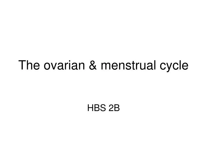
The ovarian & menstrual cycle. HBS 2B. Cell division. What type of division is shown here – meiosis or mitosis? How can you tell? Name the stages shown in each diagram. Is this likely to be happening in a male, a female or people of either sex? How can you tell?. Cell division.

E N D
Cell division • What type of division is shown here – meiosis or mitosis? • How can you tell? • Name the stages shown in each diagram. • Is this likely to be happening in a male, a female or people of either sex? • How can you tell?
Cell division Metaphase 1 Telophase 1 Prophase 1 Anaphase 1 Prophase 2 Metaphase 2 Anaphase 2 Telophase 2 • What type of division is shown here – meiosis or mitosis? • How can you tell? 2 divisions & 4 cells are produced • Name the stages shown in each diagram. • Is this likely to be happening in a male, a female or people of either sex? • How can you tell? 4 gametes are produced
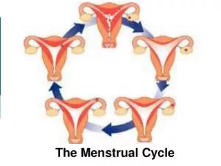
The ovarian and menstrual cycle Every month, one of the follicles in an ovary starts to develop. Each follicle consists of an immature ovum surrounded by a layer of follicular cells. As it develops the follicle gets larger and fills with fluid. After 10 –14 days the mature follicle bursts, expelling the ovum towards the uterine tube. This is called ovulation. The follicular cells develop into a structure called the corpus luteum. The ovum is wafted by movement of the fimbriae into the uterine tubes. Movement of cilia and muscular folds in the tubes cause the ovum to travel down the tubes towards the uterus. Fertilisation takes place in the uterine tubes. If fertilisation takes place then the zygote will divide and move down into the uterus where it will implant, resulting in pregnancy. If fertilisation does not take place, then the uterus will shed its lining (menstruation) and a new cycle will begin
Control of the cycle Follicle Stimulating Hormone (FSH) Luteinising Hormone (LH) Oestrogen Progesterone The reproductive system is controlled by special chemicals called hormones. Hormones are chemicals released into the blood stream that control the activity of target cells (cells with receptors for that hormone) They are made in endocrine glands, which are collections of cells that secrete into body cavities or blood stream They act by travelling in the blood until they reach a target organ, where it causes a change in activity.
Reproductive hormones The main endocrine glands associated with reproduction are the hypothalamus, pituitary gland, ovaries and testes. The placenta in a pregnant woman also acts as an endocrine gland. The hypothalamus is located at the base of the brain and is important because it controls many of the body’s activities as well as the ovarian/menstrual cycle The pituitary gland is located just below the hypothalamus and is important because it secretes many hormones that control the body’s activities – including the gonadotrophins that control the ovarian/menstrual cycle
Reproductive hormones 2 Hormonal regulation and gametogenesis in males is not cyclic as in females. Sperm are produced continually from puberty onwards. Males and females produce some hormones in common eg follicle stimulating hormone and luteinising hormone Each sex produces small amounts of hormones we associate with the opposite sex, but we consider some hormones to be mainly male eg testosterone, and some to be mainly female eg oestrogen and progesterone.
Cyclic control of the cycle 1. The hypothalamus controls release of gonadotrophins from anterior pituitary by the use of releasing and inhibiting factors 2. Release of Follicle Stimulating Hormone (FSH) from anterior pituitary causes the development of a follicle in the ovary 3. The developing follicle releases oestrogen, which acts on the uterus to repair the endometrium 4. High levels of oestrogen stimulate hypothalamus to release FSH IF & LH RF, which causes anterior pituitary to decrease secretion of FSH and increase secretion of LH. 5. Luteinising Hormone (LH) acts on the follicle to induce ovulation, and the development of the corpus luteum. 6. The corpus luteum secretes oestrogen and progesterone, which act on the uterus to prepare it for pregnancy (increased size and number of glands & blood vessels). High levels of oestrogen and progesterone inhibit the release of FSH RF and LH RF from the hypothalamus. 7. If no pregnancy occurs, the corpus luteum degenerates, and levels of oestrogen and progesterone fall. This causes the uterine lining to shed (a process called menstruation), and the hypothalamus to release FSH RF, thus starting a new cycle.
Hormonal control of spermatogenesis Gonadotropin-releasing hormone (GnRH) from the hypothalamus stimulates the anterior pituitary gland to secrete two hormones. Follicle-Stimulating Hormone (FSH) acts on Sertoli cells, which nourish developing sperm. Sertoli cells also secrete Inhibin, which reduces FSH secretion by negative feedback. Luteinizing Hormone (LH) acts on Leydig cells (interstitial cells), which produce androgens, chiefly testosterone. Testosterone regulates production of the brain hormones by negative feedback
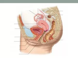
Sample test questions 1 a) Name structures: A G B H C I D J E K F b) Give the function of: i) E ii) F iii) G iv) I
Sample test questions 1 a) Name structures: A bladder G ovary B urethra H uterine tube C penis I uterus D vas deferens J bladder E testes K vagina F scrotum b) Give the function of: i) E makes sperm and testosterone ii) F protects testes and maintains them at a cooler temperature for spermatogenesis iii) G makes ova and female hormones iv) I protects and nourishes the embryo/foetus during pregnancy
Sample test questions 2 1. Look at the diagram above: Sperms A and B carry different sex chromosomes: A has an X chromosome B has a Y chromosome • What sex chromosome will the ovum contain? (1 mark) (b) If sperm A fertilises the ovum, what sex will the baby be? (1 mark) (c) What is the total number of chromosomes in sperm B? (1 mark) (d) As far as the sex chromosomes are concerned, why are there 2 sorts of sperms, but only one type of ova? (3 marks)
Sample test questions 2 1. Look at the diagram above: Sperms A and B carry different sex chromosomes: A has an X chromosome B has a Y chromosome • What sex chromosome will the ovum contain? X (1 mark) (b) If sperm A fertilises the ovum, what sex will the baby be? female (1 mark) (c) What is the total number of chromosomes in sperm B? 23 (1 mark) (d) As far as the sex chromosomes are concerned, why are there 2 sorts of sperms, but only one type of ova? Females are XX, so can only give the ovum an X chromosome. Males are XY, so will produce 2 types of sperm – half will have the X and half the Y (3 marks)