Download
1 / 30
3.87k likes | 25.87k Vues
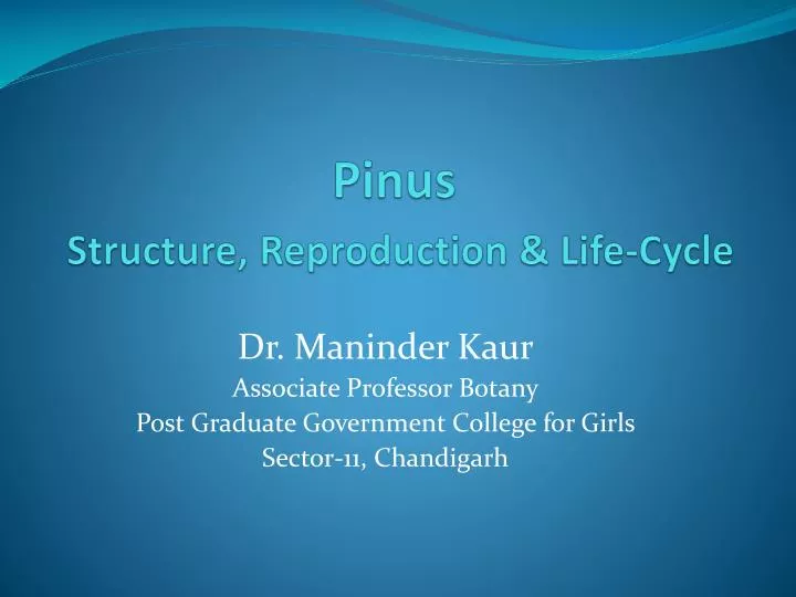
Pinus Structure, Reproduction & Life-Cycle. Dr. Maninder Kaur Associate Professor Botany Post Graduate Government College for Girls Sector-11, Chandigarh. Systematic Position. Gymnospermae Division: Coniferophyta Class: Coniferopsida Order: Coniferales Family: Pinaceae

E N D
PinusStructure, Reproduction & Life-Cycle Dr. ManinderKaur Associate Professor Botany Post Graduate Government College for Girls Sector-11, Chandigarh
Systematic Position • Gymnospermae • Division: Coniferophyta • Class: Coniferopsida • Order: Coniferales • Family: Pinaceae • Genus: Pinus
Occurrence & Distribution • Widely distributed in the Northern Hemisphere • Of the 75 species, 6 species are Indian • P. excelsa, P. longifolia, P. gerardiana, P. insularis, P. armandi • Forms dense evergreen forests in hilly regions (Himalayas)
Sporophytic Plant Body • Adult plants are tall trees up to 200 feet in height • Perennial, xerophytic plants appearing pyramidal or conical due to radial branching • Branches are dimorphic – long shoots and dwarf shoots (spurs) • Leaves are dimorphic – Scale leaves and green acicular leaves • Male and female cones present on the same plant, hence monoecious
External Morphology – Stem • Erect, tall, cylindrical, woody and branched • Branching monopodial and excurrent • Lower branches longer and horizontal giving the conical shape to the plant Pinus -trunk
External Morphology – Stem • Branches of unlimited growth are the long shoots • Arranged spirally around the main trunk • Bear scale leaves and dwarf shoots in axils of scale leaves • Branches of limited growth or dwarf shoot lacks apical bud • Possess 8-10 spirally arranged scale leaves terminating into 1-5 needle like foliage leaves at apex
External Morphology – Leaves • Scale leaves thin, brown and small • Main function is to protect young buds & conserve water around the branches • Foliage leaves are long & acicular (needle like) • Remains green for a number of years (3-10 yrs) hence plants are evergreen • No. of needles per spur varies from 1-5 with species (monofoliar to pentafoliar)
External Morphology – Root • Plant possesses tap root • Elongated structure with strong lateral branches • Root-hairs scanty; function taken up by ectotrophic mycorrhiza (fungus roots) • It is symbiotic association of fungal mycelium on the root’s surface • Helps in absorption of nutrients & protection from pathogens • Fungal species identified are Rhizopogon, Amanita, Boletus, Entoloma, etc. – mostly members of Basidiomycetes
Anatomy – Root • Resembles typical dicotyledonous root • Piliferousepiblema bear unicellular root hair (seen only in young roots) • Broad parenchymatous cortex follows • Endodermis and pericycle layers seen next • Vascular tissue is radially arranged in 2-6 groups of xylem and phloem • This tissue lacks true vessels and companion cells • Resin canals present in xylem patch making it Y-shaped • Old roots show secondary growth
Anatomy – Stem • Typically dicotyledonous stem • Cuticularized epidermis encloses the lignified sclerechymatous hypodermal layer below • Inner cortex is thin walled parenchyma containing chloroplasts and resin canals • Vascular bundles are conjoint, collateral, endarch , open and form a ring • Medullary rays are narrow • Vessels in xylem and companion cells in phloem are absent
Anatomy – Stem Pinus – T.S. of Old Stem
Secondary growth in stem • Ring of vascular cambium develops • Remains active each year forming spring wood & autumn wood – annual rings • Important in dendrology for estimation of the age of the plant • Secondary medullary rays usually uniseriate • Pinus wood is dense and massive with few parenchyma cells – pycnoxylic • Cork cambium (phellogen) formed in outer cortical layer • Forms secondary cortical cells (phelloderm) towards inner side and cork (phellem) on outer side
Anatomy – Leaf • Xeromorphic • P. longifolia is trifoliar; so the needle shows triangular outline • Outermost epidermal layer has thick-walled cells which are cuticularized • Stomata are sunken • Hypodermis is sclerenchymatous
Anatomy – Leaf Pinus – T.S. Needle
Anatomy – Leaf • Mesophyll not differentiated further • These cells have peg-like infoldings of cellulose projecting in their cavities • Have a large number of chloroplasts & starch grains • Resin canals with secretory tissue present • Two vascular bundles with conjoint tissue present in the middle Resin Duct
REPRODUCTION • Takes place by means of spores –microspores (male) and megaspores (female).The plants are therefore heterosporous • The male and female cones occur on the same plant, but different branches i.e. monoecious
Male cones (Staminate cones) • Borne on the lower branches in the axils of scale leaves. • Appear in the month of January (in plains) and March (in hills) reaching maturity within 2-3 months. • Can be seen in clusters just behind the shoot – apex.
Male cones (Staminate cones) • Each cone has 60-100 spirally arranged microsporophylls • Two microsporangia are present on the underside of each microsporophyll • Development of microsporangium is eusporangiate type • Within the microsporangium, the microspore mother cells undergo meiotic divisions to form haploid microspores
Microspore (Pollen grain) • It is surrounded by a 3-layered wall • Exine heavily cuticularized on one side of the microspore • Middle layer (exo-intine) projected outwards into two large balloon-like air sacs or wings • Inner layer(intine) is very thin • On maturation the spores germinate in situ. Hence, early gametophytic development is precocious • At the time of dehiscence, huge quantities of microspores form yellow clouds around the pine forests. It’s called the “Shower of sulphur dust” Pollen grains
Female cone (Ovulate cone) • Borne on the upper branches of the tree, in axils of scale leaves either singly or in groups of 2-4. • Female cones are seen in February and get pollinated within 3-4 months • Complete maturation and seed dispersal takes place in the 3rd year of development In Longitudinal Section
Female cone (Ovulate cone) • Each cone consists of central axis bearing spirally arranged ovuliferous scales (60-70) • On young cones a small thin & leathery bract scale can be below the ovuliferous scale • Each ovuliferous scale has two ovules on its upper surface • Cone on maturity is usually cylindrical and 15-20cms in length
Megasporophyll • The ovuliferous scale is thick, large, woody & brownish structure • More or less triangular in outline – broad, terminal portion is apophysis with its centrally projected area – the umbo • Basal portion is narrow and bears two naked, sessile anatropous ovules on its upper surface
Ovule Structure • Micropyle of the ovule faces the central axis of the cone • The single integument is fused to the nucleus except for a short distance near the micropyle • Embedded in the nucellus ,the archesporial cell divides meiotically to form four megaspores
Female Gametophyte • The inner most functional megaspore further gives rise to the haploid female gametophyte tissue wherein the archegonia develop. • The venter of the archegonia contains the upper ventral canal cell and the larger egg cell.
Male Gametophyte • Early development takes place inside the microsporangium • Pollen grains are released at the 4-celled stage (2 prothalial, a generative cell and tube cell) • Pollination is anemophilous and pollen reach the pollen chamber of the ovule through micropyle
Male Gametophyte • Further development here, results in the formation of pollen tube which carries the two unequal male gametes to the neck of the archegonium • The released male gametes will fertilize the egg cell resulting in zygote formation • Time gap of 12-14 months is seen between pollination and fertilization
Young Sporophyte • Embryo development is meroblastic • In early stages the embryonal tier of the proembryo splits apart forming 4 apical segments each with its suspensor • Each of these terminal embryonal cell give rise to a mature embryo, thus Cleavagepolyembryony is observed
Seed Structure • Seeds are naked (not enclosed in fruit) • Seeds are winged – the latter being derived from portion of upper surface of the ovuliferous scale • Outer fleshy layer of ovule disintegrates • Testa formed from the middle stony layer • Tegmen is the inner fleshy layer of the ovule • Nucellus is almost consumed during embryo development. • Remnants of nucellus , at micropylar end can be seen as reddish papery structure – the perisperm
Seed Structure • The haploid female gametophyte surrounding the embryo forms the oily white kernel (edible part). • Mature embryo has the radicle towards the micropyle and plumule away from it. • Plumule is surrounded by 8-14 cotyledons, which are green in colour. • Germination is epigeal.