Download
1 / 46
480 likes | 668 Vues
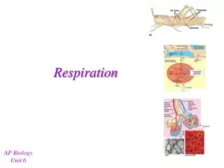
Respiration. An Introduction. I. Respiratory System Responsibilities. Removing waste product of cellular respiration (CO 2 ) Taking in gas (O 2 ) necessary for ATP formation via electron transport chain. II. Divided into four areas. Anatomy, Alveoli Structure/Function.

E N D
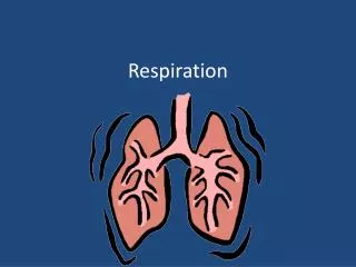
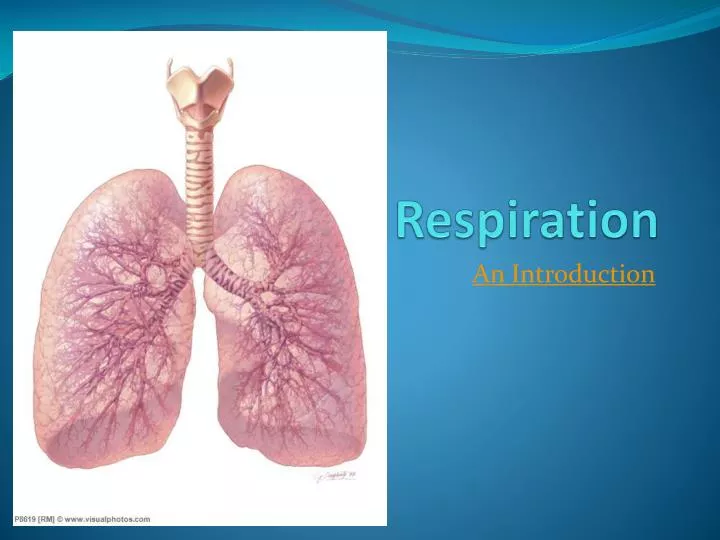
Respiration An Introduction
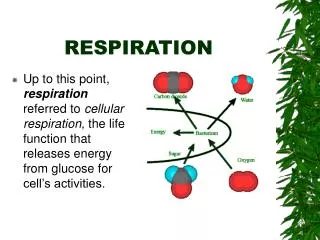
I. Respiratory System Responsibilities • Removing waste product of cellular respiration (CO2) • Taking in gas (O2 ) necessary for ATP formation via electron transport chain
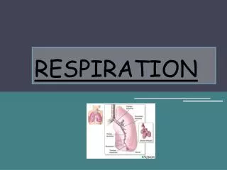
Anatomy, Alveoli Structure/Function I.Anatomy of Breathing
A. Nasal Cavity 1. NOSE contains two nasal cavities which contains narrow canals with convoluted lateral walls that are separated from one another by a septum 2. Contains special ciliated cells that act as scent receptors located at the top recesses of the nasal cavities
3. Nasal cavities are connected by tubes to the tear ducts and to the ears via the eustachian tubes 4. Air enters the nasal passages a. Air is filtered by the hairs and cilia that trap dust and debris b. Air is warmed c. Air is moistened
B. Pharynx 1. Common passage for respiratory and digestive systems 2. Above the epiglottis 3. When swallowing food, the epiglottis covers the glottis.
C. Glottis/Larynx 1. Located just below the epiglottis 2. The glottis is the opening to the larynx
3. The LARYNX is the structure that contains the vocal cords and voice box • The air enters the larynx • It is like a triangular box with the Adam's Apple at the front corner • Elastic ligaments called vocal cords stretch from the back to the front of the larynx just at the sides of the glottis • These cords vibrate when air is expelled past them through the glotti
This vibrations produce sound • The pitch of the voice depends on the length, thickness, and degree of elasticity of the vocal cords and the tension at which they are held • Muscles adjust the tension of the chords to produce different sounds
D. Trachea 1. The “windpipe” 2. Cartilaginous ridges stiffen to prevent collapse with inhalation 3. Lined with ciliated mucous membranes. a. Cilia beat upward to move up mucus and any dust or particles that were inhaled or accidentally swallowed b. Smoking can destroy cilia
E. Bronchi 1. The trachea divides into two bronchi, which branch into many smaller passages called bronchioles that extend into the lungs.
F. Bronchioles 1. The bronchioles continue to branch out, and as they do, their walls get thinner and diameter smaller 2. Each bronchiole ends in sacs called alveoli, which fill up much of the lungs
G. Alveoli Approximately 300 million alveoli per lung, for a total of 150 m2 of alveolar area (at least 40 times the area of the skin) Each alveolar sac is enclosed by a single layer of simple squamous epithelial tissue, which is surrounded by capillaries carrying deoxygenated blood
Gas exchange (CO2 and O2) diffuse directly through the walls between the blood and air in alveoli • Alveoli surfaces are moist and coated with surfactant (a lipoprotein) to prevent them from collapsing when air leaves them to reduce H2O surface tension and prevent sides from sticking to each other
H. Lungs • 1. Lungs are cone-shaped organs that lie on both sides of the heart in the thoracic cavity • 2. Branches of the pulmonary arteries follow the bronchial tubes and form a mass of capillaries around the alveoli
3. The right lung has 3 lobes and the left lung has 2 lobes 4. A lobe is divided into lobules, each of which has a bronchiole serving many alveoli 5. Lungs are very light and would float in water because they have so much air space
I. Ribs 1. Bones hinged to the vertebral column and sternum 2. Along with associated muscle, define the top and sides of the chest cavity
J. Diaphragm 1. Breathing is powered by this thick, dome-shaped muscle on the floor of the thoracic cavity (chest cavity) 2. This sheet of muscle separates the chest cavity from the abdominal cavity
K. Pleural Membranes 1. Lungs are enclosed by two pleural membranes a. Outer pleural membrane sticks closely to the walls of the chest and the diaphragm b. Inner pleural membrane is stuck to the lungs 2. The two lie very close to each other 3. In between is fluid to make for an air-tight seal 4. Pressure between the two is less than outside air pressure (or else the lung collapse when a puncture wound occurs)
L. Thoracic Cavity 1. A “sealed” chamber 2. Contains lungs, heart 3. Ribs form top and sides 4. Diaphragm forms bottom 5. Used to perform inspiration and expiration
Roles of Cilia and Mucus I. Location A. Line the tubes of the respiratory tract B. The tubes also produce mucus II. Function A. Mucus traps bacteria and dust particles B. Cilia sweep the mucus upward, cleaning the respiratory tubes C. Smokers lose functionality of this system and must cough to clear mucus (smoker’s hack)
Inhalation and Exhalation I. Breathing A. The taking in of air into the lungs 1. INSPIRATION - breathing air in 2. expiration- breathing air out B. Occurs about 14-20 times/min at rest
II. Lung Capacity A. Tidal volume 1. Normal breath 2. About 500 mL B. INSPIRATORY RESERVE VOLUME 1. Amount of air that can be forced in a breath 2. About 3100 mL
C. Inspiratory capacity 1. Maximum inhalation 2. inspiratory reserve + tidal volume • About 3600 mL D. EXPIRATORY RESERVE VOLUME 1. After normal exhalation • Can expel about 1200 mL more E. VITAL CAPACITY 1. Maximum amount of air that can be moved in and out during a single breath 2. About 4800 mL
F. RESIDUAL VOLUME 1. Air that remains in the lungs even after very deep breathing 2. About 1200 mL G. LUNG CAPACITY 1. The total amount of air in the lungs 2. About 6000 mL
III. Sequence of Events A. Breathing 1. Created by “negative pressure” powers breathing 2. Negative pressure is air pressure that is less (756 mm Hg) than the pressure of the surrounding air (760 mm Hg) 3. This negative pressure is created by increasing the volume inside the thoracic cavity 4. Air will naturally move in to fill this partial vacuum
5. The space in the thoracic cavity is made bigger by the contraction of the diaphragm and rib muscles a. Diaphragm moves downward and become less dome shaped b. When the diaphragm contracts, the space within lungs increases c. The muscles attached to the ribs, called intercostal muscles, will also contract when you breathe in d. This contraction pulls the ribs up and out, further increasing the space within the thoracic cavity 6. The decrease in the volume of the thoracic cavity forces air out of the lungs a. The diaphragm relaxes and moves upwards b. The intercostal muscles relax and the ribs move down and inward
B. Inspiration – breathing air IN 1. Diaphragm contracts 2. Rib muscles contract 3. These actions EXPAND the thoracic cavity 4. Create low pressure 5. Air is "sucked" or pulled in 6. An ACTIVE process (requires energy)
C. Expiration – breathing air OUT 1. Diaphragm relaxes 2. Ribs relax 3. Thoracic cavity relaxes 4. These actions CONTRACT the thoracic cavity 5. Air is forced out 6. A PASSIVE process
Control of Breathing I. Can be Controlled Consciously II. Mainly by Carbon Dioxide A. The urge to breathe is brought about primarily by CO2 and H+ ions in the blood 1. CO2 levels in the blood will increase as cells continue to produce it 2. The concentration of CO2 will increase until they reach a threshold level 3. High CO2 stimulates breathing center which stimulates diaphragm and rib muscles to contract
B. Chemoreceptorsin arteries detect the increased CO2 and H+levels C. The chemoreceptors send a signal to a breathing center in the medulla oblongataof the brain 1.It detects the rising levels of CO2 and H+. 2.This center is not affected by low oxygen levels.
D. There are also chemoreceptors in the carotid bodies 1. Located in the carotid arteries and in the aortic bodies, located in the aorta 2. Respond primarily to H+ concentration 3. Also to the level of carbon dioxide and oxygen in the blood 4. These bodies communicate with the respiratory center
E. The medulla oblongata sends a nerve impulse to the diaphragm and muscles in the rib cage F. The diaphragm contracts and lowers, while the rib cage moves up G. Air flows into alveoli, and the alveolar walls expand and stretch H. Stretch Receptors in the alveoli walls detect this stretching
I. Nerves in alveoli send signal to brain to inhibit the medulla oblongata from sending its message to the diaphragm and rib muscles to contract J. They therefore stop contracting K. The diaphragm relaxes, and moves upward, resuming its original shape L The rib cage relaxes and moves downward and inward M. Air is forced out the lungs
Gas Exchange I. External Respiration A. Gas exchange between AIR (at alveoli) and BLOOD (in pulmonary capillaries). B. Both alveoli walls and capillary walls are one cell layer thick. C. This exchange of gases is by diffusion alone
D. Law of diffusion states that material will flow from area of high concentration to area of low concentration E. High CO2Low CO2= conc. gradient (blood) (air – 0.5%) High O2Low O2 (air – 18%) (blood)
II. Internal Respiration A. Gas exchange of O2 and CO2 between BLOOD and TISSUE FLUID B. Oxygen diffuses from the systemic capillaries (blood) into tissue fluid HbO2 -----> Hb + O2
C. Tissue fluid is low in O2, high in CO2, due to constant cellular respiration D. CO2therefore diffuses into the blood. E. High CO2 Low CO2 (tissues) (blood) High O2 Low O2 (blood) (tissues)
Transport of CO2 and O2 in the Blood I. Oxygen A. 5% is dissolved in plasma B. 95% of blood O2 volume is oxyhaemoglobin (HbO2) II. Hemoglobin A. Hemoglobin is an iron-containing respiratory pigment found within red blood cells B. There are about 200 million hemoglobin molecules per RBC.
C. Hemoglobin increases the oxygen carrying capacity of blood by 60X D. Hemoglobin is composed of 4 polypeptide chains (a "tetramer") connected to 4 heme groups (contain iron) E. The iron portion forms a loose association with O2 F. Four O2 bind per hemoglobin molecule
G.How does hemoglobin work? • 1. Hb will bind O2 in the lungs, and release it in tissues. • 2. Hb accepts O2 more easily at cooler, more basic or neutral pH environment of lungs • 3. Hb gives up O2 at warmer, more acidic environment of the tissues
III. CO2 A. 9% dissolved in plasma • B. 27% is carbaminohemoglobin (HbCO2) • 64% combines with H2O to form carbonic acid, which dissociates in to bicarbonate ion (HCO3-) & hydrogen ions (H+) • CO2 + H2O H2CO3 H+ + HCO3- • (to Hb) ( to Plasma)
D. As CO2 levels rise due to cellular respiration, so does the H+ concentration of blood (pH drops) 1. Hemoglobin combines with the excess H+ that this reaction produces 2. Blood pH remains constant 3. Hemoglobin acts like a buffer! E. Carbonic anhydrasein RBC H+ + HCO3- H2CO3 H2O + CO2 (in blood) (to alveoli) The above reaction is driven to the right as CO2 leaves the blood, and is sped up by the enzyme carbonic anhydrase in red blood cells