Download
1 / 38
550 likes | 1.87k Vues
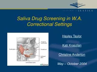
Forensic Biology Screening Workshop Saliva. Saliva . Colorless fluid secreted by 3 glands in the mouth Sublingual, submandibular, and parotid Saliva from parotid glands contain amylases, enzymes, which aid in the digestion of carbohydrates Saliva is composed of electrolytes, enzymes, mucus.

E N D
Saliva Colorless fluid secreted by 3 glands in the mouth • Sublingual, submandibular, and parotid • Saliva from parotid glands contain amylases, enzymes, which aid in the digestion of carbohydrates • Saliva is composed of electrolytes, enzymes, mucus
Saliva Screening for saliva is based on detection of high levels of amylase in the sample • It is not a confirmatory test; amylase is found in other body fluids • Serum, urine, sweat, lip mucous, semen, feces, etc. • The concentration of amylase in saliva is variable among individual; if amylase is not detected in a sample it does not mean saliva is not present
Saliva • UV light can be used to aid in locating saliva stains • The intensity of the fluorescence can be affected by the substrate, concentration of the stain, and other body fluids • Saliva does not fluoresce as intensely as semen
Saliva • Humans have both pancreatic and salivary amylase • Salivary amylase is a hydrolytic enzyme • Salivary amylase is also referred to as ptyalin
Saliva • -amylase • Found in humans elephants, rats, and pigs • Cleaves starch at its internal bonds (acts on 1,4 glucosidic linkages) allowing for compound hydrolysis-- the total breakdown of starch to maltose or glucose and dextrin • β-amylase • Found in plants • Cleaves only 1,4 glycosidic links • Can’t cleave starch at its internal bonds so there is an incomplete starch breakdown (maltose)
Amylase • One of the earliest tests for amylase was the starch-iodine test • Iodine solutions cause starch to turn a deep blue color • Amylase is a starch hydrolyzing enzyme • The presence of amylase causes the disappearance of the blue color (due to hydrolysis of the starch) and can be used an indicator for the presence of amylase
Saliva • Both α and β-amylase react with starch iodine tests • β-amylase results in a hazy/cloudy clearing due to partial breakdown of starch • α –amylase has full clearing due to complete breakdown
Amylase Testing Limitations: • Not confirmatory • Not specific for human amylase • Is specific for α-amylase • α-amylase can be found in other species
Starch Iodine Radial Diffusion Test HOW TO MAKE: • Phosphate buffer, pH 6.9 : • NaH2PO4 , anhydrous 2.7g • Na2HPO4 , anhydrous 3.9 • NaCl 0.2g • Dissolve in 500 ml of distilled water • Iodine development solution • KI 1.65 g • I2 2.54 g • Distilled water 30 ml Dissolve by stirring for 5 minutes at 65°C in a fume hood. Store saturated I2 solution in dark stoppered bottle • Working solution in 1/50 dilution with distilled water
Starch Iodine Radial Diffusion Test HOW TO MAKE: • Gel test plates (2% agarose, 0.1% soluble starch) • Phosphate buffer, pH 6.9 10.0 ml • Agarose 0.2 g • Soluble starch 0.01 g • Heat to boiling and continue stirring constantly until all the agarose is dissolved. Divide gel solution and pour into 3-2" disposable plastic Petri dishes. Allow to polymerize completely. • Store gels inverted (to retard dehydration) at 4°C.
Starch Iodine Radial Diffusion Test How to perform: • Extract a small piece of stained material with 50 µl distilled water Note: run at least one positive control consisting of a known dilution of fresh liquid saliva (1/500 in distilled water) and a negative control consisting of distilled water **some laboratories add a second positive control consisting of a 1/100 dilution of fresh saliva • Punch holes in gel plate with a vacuum pipette, leaving 1.5 cm between the sample wells • Place samples to be tested in the sample wells using a pipette. Each well holds approximately 4 µl of liquid
Starch Iodine Radial Diffusion Test How to perform: • Cover the Petri dish and place in an incubator at 37°C for 6 hours or overnight • Stain the plate by pouring a 1:50 dilution of saturated iodine solution onto the surface. Rinse with distilled water • Clear circles around the wells indicate areas of amylase activity. The diameter of the clear circle is proportional to the square root of the concentration of amylase. Record the diameter and results in notes
Starch Iodine Radial Diffusion Test How to perform: • A positive test is one in which the ring size is equal or greater in size than the positive control **some laboratories assess the level if more than one positive control is run • An inconclusive result is one in which the ring size is less than the positive control but greater than the negative control • A negative result is an absence of any clear ring
Phadebas ® Test • Commercially available • Blue starch polymer • Blue dye covalently attached to the starch and upon hydrolysis a product is obtained which is colorimetrically evaluated
Phadebas ® Test HOW TO MAKE: • Phadebas® tablets (commercially available) • 0.5 M Sodium hydroxide (commercially available) HOW TO STORE: • Follow instructions on product insert
Phadebas ® Test HOW TO PERFORM: • Place a small piece of the sample material in a 10 x 75 test tube. In a second tube, place an equal-sized piece of known saliva stain as a positive control. In a third tube add no sample (negative control) • Add 1.0 ml distilled water and ¼ Phadebas tablet to each tube using clean forceps • Vortex to mix thoroughly
Phadebas ® Test HOW TO PERFORM: • Incubate at 37°C for 30 minutes • Add 0.25 ml of 0.5 M sodium hydroxide to each tube to stop the reaction • Centrifuge for 5 minutes
Phadebas ® Test Interpretation: • A transparent dark blue supernatant of equal or greater intensity than the positive control is regarded as a positive test for amylase activity • A blue color that is less intense than the positive control but darker than the negative control is considered inconclusive for presence of amylase • No blue color is considered negative for presence of amylase
Phadebas ® Test Positive test Negative test http://www.uni-wuerzburg.de/ddch/liquits/Amylasedirekt.jpg
Amylase Mapping HOW TO PREPARE: • Use one Phadebas tablet per 5ml of distilled water • Ten tablets/50ml distilled water for piece of 46x57cm Whatman 1 filter paper • Crush tablets using mortar and pestle • Mix the Phadebas tablets with distilled water and put in a sprayer Note: Since the Phadebas mixture is actually a suspension, it must be kept mixed during spraying to evenly distribute the Phadebas particles
Amylase Mapping HOW TO PREPARE: • Hang or lay flat in a fume hood pieces of filter paper to be sprayed. Use gloves and mask to prepare papers • Spray the mist evenly onto the paper. Avoid spraying too heavy so that it does not run down the paper. Approximately 10ml per 900 cm2 gives a suitable covering • The paper can be used immediately or can be used dry Note: If papers are made up ahead of time, stored in a dark dry place
Amylase Mapping HOW TO PERFORM: • If the paper is used right away, there is no need to rewet the paper. If paper is dry, re-spray the paper with distilled water until it is damp • Caution: When rewetting, if paper is sprayed too much or too hard the blue particles will puddle • The paper is then laid spotty side down on the item under examination and its position marked • A piece of plastic sheeting is laid over the filter paper and pressed for 40-60 minutes (30 minutes at 37C) • Use of a flat board or sheet of glass on top of sheeting can aid in this process
Amylase Mapping HOW TO PERFORM: • After incubation, the paper is removed and dried. Positive areas appear as pale blue zones in place of the mottled blue negative areas. Slight shrinkage of the filter paper may occur during drying NOTE: The paper (while still damp) can be sprayed with acid phosphatase mapping reagents to search for semen stains.
SALIgAE® Test • Test available from Abacus Diagnostics • Sensitive, simple, and reported to be more accurate than other tests • Solution in a tube • Changes color with addition of an extract containing saliva • Exact mechanism – proprietary • Sensitivity - ~1:1000
SALIgAE® Test HOW TO MAKE: • SALIgAE ® Test kit (commercially available) HOW TO STORE: • Follow instructions on product insert
SALIgAE® Test HOW TO PERFORM: • Place approximately 5 mm2 cutting or ½ of a swab into a sterile 1.5 ml microcentrifuge tube • Place an equal-sized piece of known saliva stain as a positive control in a separate sterile 1.5 ml microcentrifuge tube • In a third tube add no sample (negative control) • Pipette 30 µl – 50 µl of sterile deionized water into the tube
SALIgAE® Test HOW TO PERFORM: • Incubate for 30 minutes at room temperature • Allow the test vials to warm to room temperature • Remove bubbles from the test vials by gently tapping the vials • Add 8 µl of sample to the test vial • Mix gently
SALIgAE® Test HOW TO PERFORM: • Read the result after 10 minutes • A yellow color change indicates a positive result • No color change indicates a negative result • A negative result indicates that there is no saliva present or is below the limit of detection of the test.
SALIgAE® Test http://www.dnalabsinternational.com/SalivaValidation.pdf
Rapid Stain Identification (RSID®) of Saliva • Reaction: • “S” area – antihuman salivary amylase mobile monoclonal antibody conjugated with colloidal gold • If human salivary amylase is present in the sample, a mobile Antibody-Antigen complex is formed and travels to the “T” area • “T” area - antihuman salivary amylase stationary monoclonal antibody
Rapid Stain Identification (RSID®) of Saliva • Reaction: • Stationary antibodies in the “T” area catch the mobile Antigen-Antibody complex • Forms Antibody-Antigen-Antibody complex • When the antibodies aggregate, a red line is formed • “C” area is internal control • Binds mouse antibodies with anti-mouse IgG which ensures the fluid transported the length of the strip and the test is working properly
Rapid Stain Identification (RSID®) of Saliva • HOW TO PERFORM: • Place a small cutting of the stain into a 1.5 ml microcentrifuge tube • Add 200-300 µl RSID Extraction Buffer • Incubate for 1-2 hours at room temperature • Remove 20 µl of extracted sample and add it to 80 µl of RSID TBS Running Buffer in a new tube • Add the total 100 µl to the sample well of the RSID® card • Read result after 10 minutes
Rapid Stain Identification (RSID®) of Saliva • HOW TO PERFORM: • Positive result • Test line and control line are both present • Negative result • Only control line is present • Invalid result • No line at all or only test line is present (control line absent)
Rapid Stain Identification (RSID®) of Saliva • LIMITATIONS: • False positives with breast milk, fecal material, and vaginal fluid • False negative – high dose hook effect • Sample containing up to 50 µl saliva did not result in high dose hook effect • If high dose hook effect is possible, dilute using 1:100 dilution of sample