Download
1 / 5
110 likes | 461 Vues
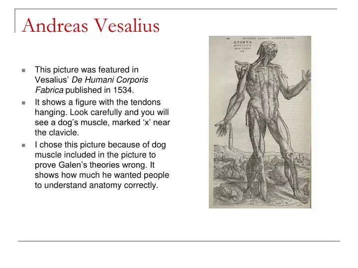
This picture was featured in Vesalius’ De Humani Corporis Fabrica published in 1534. It shows a figure with the tendons hanging. Look carefully and you will see a dog’s muscle, marked ‘x’ near the clavicle.

E N D
This picture was featured in Vesalius’ De Humani Corporis Fabrica published in 1534. It shows a figure with the tendons hanging. Look carefully and you will see a dog’s muscle, marked ‘x’ near the clavicle. I chose this picture because of dog muscle included in the picture to prove Galen’s theories wrong. It shows how much he wanted people to understand anatomy correctly. Andreas Vesalius
This drawing’s date of creation is unknown, but it was published in 1510 (after da Vinci’s death). This is a drawing of a fetus inside the uterus. I chose this picture due to the fact that it was one of the first recorded drawings of the fetus, and because of the emotions that arise when it is looked at. The concept of life is so amazing and this picture represents it very well. Leonardo da Vinci
This picture was found in a book Anatomical Engravings which unfortunately was not published until 1714. It shows a brain, the spinal cord, the many nerves attached to it. I chose this picture because it was the most accurate drawing of origin, course, and distribution of the nerves that was ever shown at that point. Bartolomeo Eustachi
This was featured in Gray’s Anatomy of the Human Body, published 1858 It is a picture of the optic nerve. I chose this picture because it reminded me of Gray’s past achievements. (In 1848, he won the triennial prize of the Royal College of Surgeons for his essay about the distribution of nerves to the human eye and it’s appendages, complete with pictures of animal’s eyes. Henry Gray
Bibliography • http://en.wikipedia.org/wiki/De_humani_corporis_fabrica • http://www.gfmer.ch/International_activities_En/Leonardo_anatomical_drawings.htm • http://www.nlm.nih.gov/exhibition/historicalanatomies/Images/1200_pixels/Eustachi_t18.jpg • http://www.bartleby.com/107/illus773.html