Download
1 / 55
560 likes | 1.26k Vues
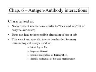
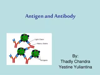
Chapter-5 Antigen antibody interactions. The reaction between antigen and serum antibodies termed serology . The interaction between Ag and Ab is the base for many immune assays, so this phenomena is used in laboratories diagnostic procedures.

E N D
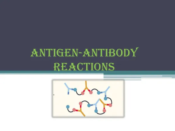
Chapter-5 Antigen antibody interactions • The reaction between antigen and serum antibodies termed serology. • The interaction between Ag and Ab is the base for many immune assays, so this phenomena is used in laboratories diagnostic procedures. • The assay may used to detect either antigen or antibody. • Serotyping of various microorganisms by using specific antisera is another application for serology. • Interaction between Ag and Ab may cause precipitation (in case of soluble Ag) or agglutination (in case of solid Ag). • Agglutination and precipitation need multivalent antigen and antibody with at least TWO binding sites.
Chapter-5 Antigen antibody interactions Unideterminant and Univalent Unideterminant and Multiivalent Multideterminant and Multivalent
Chapter-5 Antigen antibody interactions • For precipitation, agglutination and complemet activation Cross-linking of various antigens should take place. + = + =
Chapter-5 Antigen antibody interactions Primary interactions between Ab and Ag: • Binding forces between Ab and Ag can be Hydrophobic force, electrostatic force or Van der Waals force. • The bonding strength between Ab and a univalent Ag termed as affinity. Whereas the term avidity is overall binding strength between Ab and multivalent antigen. Secondary interactions between Ab and Ag: - Cross-linking of many antigens by antibodies cause agglutination or clumping of antigens.
Biology of the B-lymphocytes • B cells are cells that secret antibodies. • B cells acquired their name from early experiments in birds (gland called bursa). • B cells development depends upon Ig gene rearrangement. • B cell development go through different stages. Stem cells Pro-B Pre-B Immature-B Memory Mature-B Sites of early B cell differentiation: • During fetal development (fetal liver). • After birth mainly in bone marrow.
Biology of the B-lymphocytes • Cytokines play a role in the early proliferation and differentiation of haematopoietic stem cells. • As a result of rearrangement in the V(D)J region of the Ig gene, Pro-B cell is the first cells to be generated. Rearrangement takes place in the heavy-chain locus (DH gene segment to JH segment). • Pro-B cell express CD19 and CD10. • The next step of B cell differentiation is Pre-B cell. The heavy chain VH gene segment rearranges and join DHVH segments and VDJ then formed. • The new rearrangement (VDJ unit) is close to another region called C. segment is the nearest to the VDJ unit. • Pre-B cell synthesise chain. At this stage express CD19 and CD10.
Biology of the B-lymphocytes • chain expressed as a transmembrane molecule. • On the membrane found in conjunction with products of two genes 5 and VpreB. • 5 and VpreB together called surrogate light chain. • 5 and VpreB products are Ig and Ig which both linked by disulfide bond. • The chain and the surrogate light chains is referred to as Pre-B-cell receptor (pre-BCR). • Ig and Ig are responsible for signal transduction in mature B-cells (signal transduction molecules). • In Pre-B cell the Ig and Ig. and through the pre-BCR a signal a stop signal in the heavy chain gene rearrangement and L-chain gene rearrangement.
Biology of the B-lymphocytes • Rearrangement in L-chain gene starts with -chain genes then the -chain genes. • Rearrangement in L-chain genes involves only V and J genes. • The signal given by the (Pre-BCR) cause differentiation of the cell to next stage. • The signal caused by the Pre-BCR need to reach the nucleus and this involves a specific enzyme (Bruton’s tyrosine kinase). • Some individuals lack (Btk) due to mutation in the gene responsible for coding the enzyme causing NO differentiation for the Pre-B-cell. This lead to the development of an immuno-deficiency condition know X-linked agammaglobulinemia. • After the synthesis of and L chains they pair together to form monomeric IgM which appears on the surface of the cell
Biology of the B-lymphocytes • Cell at this stage called immature B cell. • At this stage B-cell can recognise ONLY self antigens (presented by MHC molecules on bone marrow cells). • This interaction activates a phenomena called receptor editing. • Receptor editing involves a secondary rearrangements in the V(D)J region and mainly in the light chain ( and loci). The rearrangement takes place in the unrearranged genes during the primary V(D)J rearrangement. • The overall rearrangement process lead to the generation of Ig specificity either for SELF ANTIGEN or NON-SELF ANTIGEN. • If B-cell with SELF ANTIGEN generated then the immature B-cell deleted (apoptosis) or become anergic.
Biology of the B-lymphocytes • Survived cells then differentiate into mature cells after express IgD on their surface and become IgM+ and IgD+. • Plasma cells is the antibody secreting cells. They lack CD19, CD10 and CD20. • Memory B cells : They express Igs like IgG and IgE on their receptors beside IgM but not IgD. They also express CD27 as a marker. B-CELL MEMBRANE PROTEINS: 1- Antigen binding receptors Igs receptors (IgM and IgD). Pro-B cell and plasma do NOT express these binding receptors. 2- Signal transduction receptors associated with membrane Igs, these include Ig and Ig (Pre-BCR). The cytoplasmic tail has immunoreceptor tyrosine based activation motifs (ITAMs).
Biology of the B-lymphocytes 3- B-cell coreceptors, CD19, CD81 and CD21. Such receptors augment B-cells response to a particular Ag. 4- Negative regulation of B-cell signalling, a receptor like CD22 has a negative regulating effect on CD19, CD21, CD81 and CD32 (Fc-IgG-III). 5- Receptors involved in T-cell B-cell interactions, these receptors include MHC-II (stimulatory receptor). A costimulatory molecules needed beside Ag to activate T-cells. Costimulatory molecules include B71-2(CD80 and CD86) and CD40. CD40 interaction with its counter ligand on T-cell (CD40L or CD154) has an important role in Ig switching. X-linked hyper-IgM syndrome is related to mutation in CD154 gene so B-cells can not undergo Ig isotype switching (they produce only IgM).
Biology of the B-lymphocytes • Inducible-costimulatory (ICOS) ligand (ICOSL) is important for T-cell B-cell interaction, ICOSL on B-cells may interact with ICOS expressed on activated T-cells. Individuals lack ICISL or ICOS have low concentration of IgG, IgA and IgE. B-CELL ADHESIVE(ADHESION) MOLECULES: Such receptors include CD54 (ICAM-1), CD58 (LFA-3). Biology of the T Lymphocytes- Chapter 9 • T-cell is the cell that recognise Ags. • T-cell recognise Ag through a specific receptor called TCR.
Biology of the T-lymphocytes • T lymphocytes development takes place in the thymus. • MHC molecules play an important role in the development of T lymphocytes in the thymus. • Acquiring TCR is essential for the development of T-cells. • Every clone of T-cells express a TCR with unique sequence (specific for one antigenic determinant or epitope). TCR: • It is a complex made up of more than one part. One part is variable and responsible for Ag binding and other parts which are invariant and used for signal transduction. Molecules that bind Ags (variable): • Two polypeptide chains termed and , they are transmembrane glycoproteins with short cytoplasmic tails.
Biology of the T-lymphocytes • Both and has two domains one is variable (V and V) and the other is constant (C and C). • TCR V and V has three complementarity-determining regions CDR1, CDR2 and CDR3). • Although TCR is very similar to Igs structure (Ig superfamilly) yet they are different in many aspects: ** Ig is divalent whereas TCR is monovalent. ** Ig has hinge region which helps him binds all types of Ags (soluble or surface). TCR is rigid with no hinge regions it only can binds Ages on cell surfaces. ** Ig can bind all classes of Ags (proteins, carbohydrates, lipids and DNA), whereas TCR binds small fragments of proteins. ** Igs secreted post activation but not TCR. ** Igs change (isotype switching) TCR do not.
Biology of the T-lymphocytes Invariant molecules of TCR: • Found noncovalently associated to and chains. CD3 and two identical chains (zeta or CD274). • CD3 consists of three polypeptide chains , and (each has one domain). • CD3 chains contains tyrosine sequence (immunoreceptor tyrosine-based activation motif). Coreceptors molecules: • Coreceptors in T- cells are transmembrane molecules and very similar in function to those coreceptors found on B-cells. CD4 and CD8 are T-cells coreceptors. • CD4 and CD8 bind the invariant part of the MHC molecules.
Biology of the T-lymphocytes • CD4 and CD8 also act as adhesion molecules. • Because CD4 and CD8 are linked to protein tyrosine kinases they involve in signal transduction. Costimulatory receptors: • Naïve T-cells need costimulatory signal or interactions to be fully activated. CD28 on T-cells interact with B7-1,2 (CD80 and CD86) on APC. CD40 on T-cells interact with CD154 on APC. CD152 expressed on activated T-cells interact with B7 molecules for negative signalling on the activated T-cells. Adhesion molecules: CD2 on T-cells interact with CD58 on APC. CD62L homing and adhesion molecule
Role of major histocompatability complex in the immune response Major Histocompatability Complex (MHC). • MHC molecule plays an important role in T-cell response to Ag (also maturation of T-cells in the thymus). • T-cell response is referred to as MHC restriction of T-cell response. • MHC molecules coded by a set of genes (complex) closely located to each other. MHC genes: • In human MHC genes located on chromosome 6, and the molecule called human leukocyte antigen (HLA), in mouse it’s called MHC-H-2 and located in chromosome17. • There are TWO sets of MHC genes known as MHC class I and MHC class II.
Major histocompatability complex • MHC molecules play a role in T-cell response, MHC-I interact with CD8+ T-cells. • MHC-I molecules expressed on all nucleated cells. • MHC-I bind peptides derived from proteins found in the cytoplasm of an infected cell. • CD4+ interacts with MHC-II. • MHC-II expression is restricted and found only on the surface of phagocytic cells (APCs eg. Macrophage and dendritic cell). • Expression of both MHC molecules can be regulated by many factors like cytokines, interferons (IFN) , and . VARIABILITY OF MHC MOLECULES: • MHC I and II molecules are different from one individual to another. The variation is due to two reasons:
Major histocompatability complex I- Polygenicity. II- Polymorphism. • Polygenicity means that MHC molecules coded by more than one independent gene each (multiple). • THREE independent genes found to code for MHC-I named HLA-A, HLA-B and HLA-C. NOTE THAT THE TREE MOLECULES MAY FOUND ON ONE CELL SURFACES. • Because you have TWO homologous chromosomes (paternal + maternal) then you should have TWO sets of genes each with three genes. So the final and maximum number of different MHC-I molecules one cell can express on its surface is 32= 6 molecules. A cell can express less than six different molecules in some cases. • MHC-II also coded by THREE independent genes known as HLA-DP, HLA-DQ and HLA-DR. So again one cell can express SIX different MHC-II molecules on its surface.
Major histocompatability complex • Polymorphism of the HLA coding genes. HLA system is the most highly polymorphic gene system in human body. • Different alleles of every gene codes for MHC molecules. • MHC molecules expressed co-dominantly on surface of cells.
Major histocompatability complex MHC class I structure: • MHC-I consists of one transmembrane glycoprotein chain with three extracellular domains (1, 2, 3), the three domains associated non-covalently with another invariant polypeptide called 2-microglobulin. • 2-microglobulin coded by a different chromosome. • 1 and 2 are variant and unique to the allele coded for their synthesis. • 3is invariant into all MHC-I molecules. • Space between 1 and 2 called the peptide-binding groove where the polypeptide (Ag) going to be presented (binding site with T-cell TCR). • CD8 bind MHC-I molecule on the 3 domain, TCR of the CD8 cell binds on Ag peptide presented on the groove.
Major histocompatability complex MHC class I structure:
Major histocompatability complex MHC class II structure: • MHC-II consists of two transmembrane glycoprotein chains each with two extracellular domains (1, 2 and 1, 2). • Like MHC-I variation found in 1 and 1 and they are unique to the allele coded for their synthesis. • 2 and 2 are invariant into all MHC-II molecules. • Space between 1 and 2 called the peptide-binding groove where the polypeptide Ag going to be presented (binding site). • CD4 bind MHC-II molecule on the invariant region, TCR of the CD4 cell binds on Ag peptide presented on the groove pocket.
Major histocompatability complex MHC class II structure:
Major histocompatability complex Genes of the HLA region: • I human, MHC-I and II genes are located on chromosome 6. • For HLA-I genes products (HLA-A, HLA-B and HLA-Cw to be distinguished from complement factor C2 and C3 etc..) • The 2-microglobulin coded by a different chromosome (chromosome 15). • MHC-I genes has many alleles as much as 630 for gene A and 979 for gen B and 338 for gene C. • It is almost impossible to fined two genetically identical individuals. • In cross matching (serologically) antibodies against specific HLA-I molecules tested by giving numbers (HLA-A17, HLA-B39 and HLA-C25).
Major histocompatability complex • For HLA-II the su-bregions DP, DQ and DR each has two genes A and B. A gene code for chain and gene B code for chain. So HLA-DPA1 code for HLA-DP and HLA-DPB1 code for HLA-DP. • DR sub-region has two B genes (B1 and B2). • MHC-II genes also have many alleles. Others genes within MHC region: • MHC-I and MHC-II genes is separated by a group of genes known as MHC-III. • MHC-III genes have nothing to do with antigen presenting. • MHC-III are genes code for complement factors like C2, C4 and factor B. • HLA-E, HLA-F and G also found within the region of HLA-I is believed they involve in Ag presentation to NK cells.
Major histocompatability complex ANTIGEN PROCESSING AND PRESENTATION: Exogenous Antigens: • Antigens that found outside the host cell and usually internalised into the host by endocytosis (phagocytosis or pinocytosis). • The antigen can be any thing from bacteria or its product or even a vaccine (a dead virus). - The antigen get internalised by phagocytic cell into a phagosomal vacuole. The vacuole fused with the cell lysosome. Due to low pH and the presence of hydrolytic enzymes (proteases and lipases…) the antigen mostly degraded and become processed. - An immune determinant formed (antigenic determinant).
Major histocompatability complex - In the endoplasmic reticulum HLA synthesise( and ). Then the molecule is assembled with a small fragment called invariant chain (Ii or CD74). - CD74 found bound to the groove site of HLA molecule. - HLA/CD74 enters the Golgi apparatus and CD74 starts degraded proteolytically, a very small portion of CD74 stays untouched, this portion called CLIP (Class II associated Invariant Polypeptide). CLIP usually found blocking the groove site. - HLA/CLIP leaves the Golgi towards the phagosomal cavity where it fuses with it. Once inside an HLA-DM molecule catalyse the exchange between the CLIP bound to the HLA-II and the processed peptide (antigenic determinant). The HLA-II/peptide then moved to the surface of the cell. CD4+ cell always associated with antigens presented by HLA-II molecules.
Major histocompatability complex Endogenous antigens: • Endogenous antigens are derived or synthesised inside the host cell (mostly intracellular pathogens). • Endogenous antigens usually processed in the cytoplasmic compartment. The resulted immune determinant (7-10) amino acid then transported to the endoplasmic reticulum. The peptide transported by a transporter called TAP (Transporter associated with peptide). • In endoplasmic reticulum HLA-I synthesis and assembled ( and 2 microglobulin). The immuno domenant binds the HLA-I on the binding site. The complex then moves via Golgi apparatus to the cell surface where CD8+ cells can recognise and associate with the presented antigen.
Activation of T and B lymphocytes • APC initiates the activation of naïve T-cell. • APC interact with Ag by phagocytosis through various recognition receptors include MB (Mannan-binding lectin) toll-like receptors (TLRs), most cells involved in innate immunity express TLRs. • TLRs can be for lipoproteins, lipopolysaccharide, mannose residues. • Activation of uncommitted (resting) T-lymphocytes by an APC (e.g.. M, DC) lead to Proliferation and Differentiation of T-lymphocytes to effectors' cells. • Various types of cytokines secreted as a result. • Activation of T-lymphocytes involve both CD4+ and CD8+ T- lymphocytes.
Activation of T and B lymphocytes Dendritic cells and activation of T lymphocytes: • DC found in many tissues include ones close to secondary lymphoid organs. • Two major clones of DC (plasmacytoid, myeloid). • Plasmacytoid DC plays a role in innate immunity and secretes IFN- and whereas myeloid DC plays a role in CD4+ cell activation. • DC found in tissues referred to as immature dendritic cells. These cells can interact with different Ags by different pattern recognition receptors PRR (e.g. TLRs) which can interact with bacterial DNA, lipopolysaccharide and viral RNA. • Processing and presentation of phagocytosed Ag induces the expression of HLA-II and costimulatory molecules.
Activation of T and B lymphocytes • Array of cytokines secreted by activated DC like IL-12. • Activated DC migrate to secondary lymphoid organs where T-cell housed. Antigen presenting DC now referred to as mature dendritic cell and present the Ag to the CD4+ cell. Intercellular events in CD4+ cell activation: • The signal initiated by the TCR (variable parts and ) HLA-II interaction, a signal passed to other parts of the TCR complex (CD3 and ). The cytoplasmic portion of CD3 and involves with activated tyrosine kinases Fyn. The cytoplasmic portion of CD 4 associated Lck. Once Fyn and Lck activated by a membrane protein CD45 they bind CD3 and and cause phosphorylation of these binding regions. Phosphorylation of Fyn and Lck enable another tyrosine kinase (ZAP-70)to bind . Lck is responsible for the activation of bound ZAP-70 (individuals lack ZAP-70 do not respond to antigens).
Activation of T and B lymphocytes • ZAP-70 activates two adapter proteins (phosphorylation). • The activated adapter molecules bind phospholipase C- (PLC-). The (PLC-) activates another membrane molecule called phospholipid phosphatidylinositol biphosphate (PIP2). • PIP2 slplit into two components diacylglycerol (DAG) and inositol triphosphate (IP3). • As a final result of intracellular signalling gene expression for synthesis of particular molecules like IL-2 and CD25 is taken place. • Interaction between T-cell and APC takes place in a stretch of area called immunologic synapse. Superantigens: • Molecules that activates all T-cells with a particular V gene segment with irrespective of their V expression. These molecules mostly bacterial toxins.
Activation of T and B lymphocytes • Other co-receptors (ligands) are involved: Adhesive receptors: - CD18 (LFA-1)-------------------------- CD54 (ICAM-1) - CD2-------------------------------------- (CD58) (LFA-3) - CD29 ------------------------------------ (CD106) (VCAM-1) Co-stimulatoy receptors: - CD4-------------------------------------- (MHC-II) - CD28------------------------------------- (B7-1,2, CD80, CD86) - CD152----------------------------------- (CD80, CD86) - CD154 (CD40L)------------------------ (CD40) - ICOS--------------------------------------(ICOSL)
Activation of T and B lymphocytes Subsets of CD4+ cells: • CD4+ cells and according to cytokines released and the distinct effector cells beside the function they perform they can be divided into FOUR subsets: TH1, TH2, TH17 and Treg.
SubsetSecreted ILsAct onfunction TH1 IFN-, IL-2 M,NK, CD8 cell mediated imm. B-cell to secrete IgG3 intracellular viral & bacterial TH2 IL-4, IL-5, Eosinophil Response to worms IL-13 B-cell to secrete IgE and allergens. TH17 IL-17, IL-22 Neutrophils & Proinflammatory Epithelail cells responses to fungi extracellular. bacteria Treg IL-10,TGF- activated inhibits function of lymphocytes T-cells and others.
Activation of T and B lymphocytes CD4 T-CELL DIFFERENTATION: • Cytokines secreted by TH1 T-cell CD4+ secrete IL-2, IFN- and TNF- these cytokines known for their positive effect on CD8 and NK cells. They also have an effect on B-ells to secrete certain isotypes of immunoglobulins that activate complement. • TH1 cytokines play a role in cell mediated immunity. • Cytokines secreted by TH2 include IL-4 IL-5, IL-13. IL-4 and IL-13 play a role in B-cell class switching to IgE and IgG4. IL-5 has a role during parasitic infections through eosinophils. • TH17 T-cell CD4+ secrete cytokines IL-17 and , IL-22. These cytokines are promoting the synthesis of proinflammatory cytokines IL-1, IL-6 and TNF-. These cytokines found to have a major role during inflammation majorly at mucosal sites. TH17 cells and cytokines are involved in human autoimmune diseases e.g rheumatoid arthritis and multiple sclerosis.
Activation of T and B lymphocytes • Treg cells secret cytokines that’s suppress or regulate the function of other T-cells subsets and many cells like APCs. Treg secrets IL-10 and TGF-. Treg lackness may lead to serious immune dysregulation which could lead to autoimmune diseases and allergy. Cytokines influence differentiation of CD4+ TH0: • Signal from activated APC (e.g. dendritic cell) plays a role in the differentiation of naïve CD4+ cell (TH0). This signal resulted from the type of cytokines secreted by these APC (this also referred to as (third signal). • IL-12 secreted by NK and DC against particular bacteria and viruses induces the development of TH1. IL-4 from activated mast cells promote the development of TH2. TGF- and IL-21 promote the development of TH17 (produced by CD4+).
Activation of T and B lymphocytes Cross inhibition of CD4+ T-cells subsets: • Cytokines secreted by a particular subset inhibits the secretion of cytokines by other subsets. - IFN- from TH1 inhibits the TH2 development. - IL-4 from TH2 inhibits the TH1. - IFN- TH2 inhibits the TH17 development. - Treg cytokines inhibit the development of other T-cell subsets. • The immune response to a particular antigen is polarised.