Download
1 / 56
640 likes | 1.56k Vues
Mycology. Dental / Optometry Fundamentals II Stephen A. Moser, Ph.D. 10/26/2011. Epidemiology. Geography Endemic mycoses Worldwide mycoses Transmission of infection Respiratory inhalation (systemic mycoses) Cutaneous inoculation (sporotrichosis)

E N D
Mycology Dental / Optometry Fundamentals II Stephen A. Moser, Ph.D. 10/26/2011
Epidemiology • Geography • Endemic mycoses • Worldwide mycoses • Transmission of infection • Respiratory inhalation (systemic mycoses) • Cutaneous inoculation (sporotrichosis) • Systemic invasion by opportunistic normal flora (candidiasis) • Contact with infected hosts (dermatophytoses)
Epidemiology (Cont.) • Risk factors and manifestations of disease • True pathogens versus opportunists • Environmental risk factors for systemic fungal disease • Location and travel • Occupation • Host defenses and susceptibility to systemic fungal disease (CMI most important) • Congenital and acquired T cell deficiencies (including AIDS) • Immunosuppression (transplants and malignancies) • Diabetes mellitus
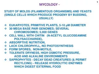
General Characteristics • Aerobic - obligate or facultative • Eukaryotic: membrane bound nucleus and cytoplasmic organelles (may be multinucleate) • Achlorophyllous • Morphology (unicellular or multicellular) • Saprophytic (heterotrophic)
Characteristics of Fungal Cells • Cell wall: multilayered polysaccharide • Cellulose, glucans, mannans, chitin, polypeptides • Absence of teichoic acids, peptidoglycan, LPS • Cell membrane • Phospholipid bilayer • Ergosterol (relate to chemotherapy) • Cytoplasm- typical eukaryotic organelles • Nucleus- either uninucleate or multinucleate
Characteristics of Fungal Cells • Capsule • Present in some species (e.G. Cryptococcus neoformans) • Amorphous polysaccharide coating • Functionsand activities • Antiphagocytic • Antigenic
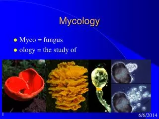
Characteristics of Fungal Cells • Growth forms • Yeast - unicellular fungi which reproduce by budding (Cryptococcus) • Mold - hyphae (mycelium) • Septate hyphae (Aspergillus) • Non-septate, coenocytic hyphae (Mucor) • Pseudohyphae (Candida albicans) • Thermal dimorphism
Examples of Yeast & Pseudohyphae Blastoconidia Pseudohypha
Asexual Reproduction • Conidia (spores) – asexual structures • Blastospores – formed by budding yeasts (Blastomyces) • Chlamydospores – terminal or intercalary cells with thick walls (Candida albicans) • Arthrospores – formed by fragmentation of hyphae (Coccidioides immitis) • Sproangiospores – formed in sporangia by cleavage (Rhizopus)
Classification Based onSexual Phase • Ascomycetes: Aspergillus,Histoplasma, Blastomyces, Dermatophytes • Basidiomycetes: Cryptococcus, Mushrooms • Zygomycetes: Order Mucorales - Mucor, Rhizopus • Deuteromycetes (Fungi Imperfecti): Sporothrix, Coccidioides, Candida
Routes of Infection • Inhalation of spores – major factor • Inoculation of spores into skin • Disease by normal flora in compromised host (Candida) • Hypersensitivity • Contact with infected host (Dermatophytes) • Mycotoxins
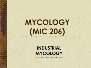
Laboratory Diagnosis of Fungal Infections • Microscopic Examination of tissues and body fluids • Gram stain • Giemsa • India Ink • Potassium hydroxide (KOH) wet prep • Hematoxylin and Eosin stain • Periodic-Acid Schiff stain (PAS) • Gomori-Methenamine Silver stain (GMS) • Mucicarmine or Alcian Blue stain
Budding Yeast - Gram Stain Staphylococcus Candida
Histopathological Response to Fungal Infection • Acute pyogenic abscess (Candida) • Chronic granuloma formation (Histoplasma) • Chronic, localized dermal inflammation (Dermatophytes) • Mixed pyogenic and granulomatous inflammation (Blastomyces) • Blood vessel invasion with thrombosis and infarction (Mucor, Aspergillus) • Hypersensitivity without tissue reaction (allergic bronchopulmonary aspergillosis)
Fungal Cultures • Utilize Sabouraud agar with antibiotics • Identification criteria • Temperature of growth • Rate of growth • Colonial and microscopic morphology • Sporulation pattern • Biochemical reactions (yeast)
Fungal Serology • Generally poor and not as useful as in other pathogens such as viruses and bacteria, with some exceptions. • Cryptococcal antigen by latex agglutination: serum and CSF. • Coccidioides - early IgM response is useful for identification of acute primary disease - CSF IgG prognostic value. • Skin tests for DTH - problems: • Cross-reactivity. • High positive rate in endemic areas.
Candidiasis • Clinical manifestations • Mucosal • Vaginitis • Esophagitis • Oral thrush • Cutaneous • Chronic mucocutaneous • Systemic • Fungemia • Hepato-spleenic • Endophthalmitis • Renal • Urinary tract
Aspergillosis • Clinical manifestations • Pneumonia • Aspergilloma • Allergic bronchopulmonary • Disseminated multiorgan involvement
Zygomycosis • Clinical manifestations • Sinusitis • Rhinocerebral • Pulmonary • Renal
Histoplasmosis • Clinical manifestations • Most cases mild or sub-clinical pulmonary disease • Dissemination appears to be common • Pneumonia • Chronic progressive pulmonary (cavitary) • Histoplasmoma • Disseminated
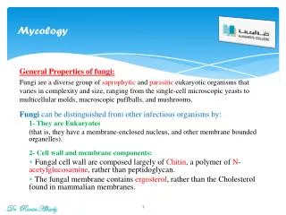
Histoplasmosis – Bone Marrow H. capsulatum Histiocyte
Histoplasma capsulatum In vitro In vivo
Presumed Ocular Histoplasmosis • Thought to be a late stage of primary histoplasmosis. • Causes abnormal blood vessels – scar tissue. • Organism has not been found in eye. • Treated with laser surgery.
Chemotherapy • FDA approved • Polyenes (Amphotericin B, lipid encapsulated forms) • Azoles (fluconazole, itraconazole, ketoconazole, voriconazole) • Echinocandin (Caspofungin, Micafungin, Anidulafungin) • Nucleoside derivatives (5-flurocytosine) • Allyamines (Terbinafine) • Microtubule disruption (Griseofulvin) • Investigational • Nikkomycins (chitin synthase inhibitors) • Echinocandin/pnemocandin/lipopeptide class (inhibit glycan synthesis)