Download
1 / 54
990 likes | 2.2k Vues
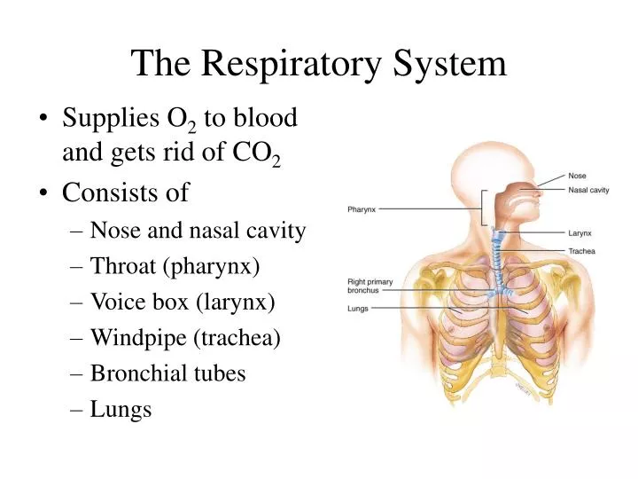
The Respiratory System. Supplies O 2 to blood and gets rid of CO 2 Consists of Nose and nasal cavity Throat (pharynx) Voice box (larynx) Windpipe (trachea) Bronchial tubes Lungs. Functions of the Respiratory System. Cleaning and filtering air Ventilation Gas exchange Gas transport

E N D
The Respiratory System • Supplies O2 to blood and gets rid of CO2 • Consists of • Nose and nasal cavity • Throat (pharynx) • Voice box (larynx) • Windpipe (trachea) • Bronchial tubes • Lungs
Functions of the Respiratory System • Cleaning and filtering air • Ventilation • Gas exchange • Gas transport • Smell • Speech
Respiratory System Anatomy • Upper respiratory system (tract) • Nose and nasal cavity • Pharynx = throat • Lower respiratory system (tract) • Larynx = voicebox • Trachea = windpipe • Bronchi = airways • Lungs • Refer to locations of infections
Trachea • Five inches long & one inch in diameter • Extends from larynx into chest where it divides to form two bronchi(us) • 16 to 20 incomplete C-shaped cartilage rings • Lined with pseudostratified epithelium Trachea--------------- Bronchus--------------------------- --------------Bronchus
Gross Anatomy of Lungs • Base, apex, cardiac notch • Oblique & horizontal fissure in right lung results in 3 lobes • Oblique fissure only in left lung produces 2 lobes
Lung Lobules • Lung lobules are smaller compartments within lobes • Consist of • Terminal bronchiole supplies air to lobule • Each terminal bronchiole divides into several respiratory bronchioles • Respiratory bronchioles divide into alveolar ducts • Alveolar ducts supply air to alveolar (air) sacs • Each sac composed of two or more smaller alveoli(us) • Extensive blood supply ---terminal bronchiole ---respiratory bronchiole alveolar duct--------------------- ---alveolar sac -------alveolus
Details of Respiratory Membrane • Four tissue layers & two fluid layers make the respiratory membrane.
Respiratory Physiology Ventilation, Gas Exchange, Gas Transport and Control of Ventilation
Pulmonary (alveolar) Ventilation • Basic concepts and definitions • Ventilation called negative draft ventilation • Breathing in called inspiration or inhalation • Breathing out called expiration or exhalation • Pressure within the lung called alveolar pressure • Pressure within the pleural cavities called intrapleural pressure – always less than atmospheric pressure. Keeps lungs attached to chest wall and inflated
Breathing or Pulmonary VentilationQuiet Ventilation - Eupnea • Air moves into lungs when pressure inside lungs is less than atmospheric pressure • Contraction of diaphragm and rib muscles (external intercostals) enlarges chest and reduces alveolar pressure to below atmospheric pressure • Air drafts into lungs – negative draft • inspiration or inhalation
Breathing or Pulmonary Ventilation • Air moves out of lungs when pressure inside lungs is greater than atmospheric pressure • Diaphragm and rib muscles (ext. Intercostals) relax • Chest gets smaller • Elastic recoil of alveoli creates Alveolar pressure greater than atmospheric pressure • Air is pushed (squeezed) out of lungs • Expiration or exhalation
Boyle’s Law and Ventilation • As size of container increases, pressure inside decreases • As the size of closed container decreases, pressure inside is increased
Forced Ventilation During Exercise • Forced inspiration • Require larger decreases in intrapleural and alveolar pressures • Diaphragm and external rib muscles contract more forcefully making the chest wider. • Other back muscles become involved • Results in deeper breaths
Forced Ventilation During Exercise • Forced Expiration • Require larger increases in intrapleural and alveolar pressures • Diaphragm and external intercostal rib muscles relax • Internal intercostal rib muscles contract compressing rib cage making the chest narrower. • Abdominal muscles contract compressing abdomen forcing diaphragm to move up further • Air forced out
Lung Volumes and Capacities • Volume is one measure of quantity of air • Capacity is sum of two or more volumes • Spirometer or respirometer device for measuring volumes and capacities • Record called spirogram
Lung Volumes and Capacities • Ventilation rate is number of breaths per minute • Resting ventilation rate averages12 breaths per minute (Range 12 to 20 breaths / minute) • Tidal Volume (VT) is amount of air in one breath • Resting tidal volume is about 500 mL
Lung Volumes and Capacities • If resting tidal volume (VT) = 500 mL, then • 350 mL reaches alveoli • Remaining 150 mL remains in conducting airways above alveoli • Called anatomic dead air • Dead air does not participate in gas exchange • The 350 mL in alveoli is the only air participating in gas exchange
Lung Volumes and Capacities • Lung volumes (1) Tidal Volume (Vt) Volume of air in one breath (2) Inspiratory Reserve Volume (IRV) Volume of air inspired in addition to VT (3) Expiratory Reserve Volume (ERV) Volume of air expired in addition to VT (4) Residual Volume(RV) Volume of air that cannot be expired even with maximum forced expiration.
Lung Volumes and Capacities • Lung Capacities: (1) Inspiratory Capacity (IC) = Vt + IRV (2) Functional Residual Capacity: (FRC) = RV + ERV (3) Vital Capacity(VC)= Vt + IRV + ERV (4) Total Lung Capacity(TLC) =VC + RV
Lung Volumes and Capacities • FEV1 stands for forced expiratory volume in one second • Percentage of vital capacity expired in 1 second • Should be 75% or higher • If not, may indicate chronic obstructive pulmonary disease such as chronic bronchitis or emphysema
Gas exchange • In lungs – External respiration • Diffusion of O2 from alveolar air into blood • Diffusion of CO2 from the blood into alveolar air • In tissues – Internal respiration • Diffusion of O2 from blood into tissues • Diffusion of CO2 from tissues into blood • Diffusion across the extremely thin respiratory membrane from higher to lower concentrations of gases
External Respiration Gas Exchange Diagram Internal Respiration O2 O2 CO2 CO2 O2 Oxygenated Blood Alveolus CO2 O2 CO2 O2 O2 CO2 CO2 Deoxygenated Blood Tissues
Measurements of Gas Concentrations • Dalton’s Law of Partial Pressure • In a mixture of gasses, the total pressure is equal to the sum of pressures contributed by each individual gas • These individual pressures are partial pressures • Symbol for the partial pressure of a gas is Pg where g stands for the specific gas
Measurements of Gas Concentrations • Partial pressures • Our atmosphere is mixture of nitrogen, oxygen, argon, carbon dioxide and other trace gasses • Total pressure of our atmosphere is essentially equal to PN2 + PO2 + PAr + PCO2
Measurements of Gas Concentrations • Partial pressures • Since Oxygen (O2) = 21% of our atmosphere, its PO2 = 0.21 x 760 = 159 mm Hg • Since CO2 = 0.04% of our atmosphere, its PCO2 = 0.0004 x 760 = 0.3 mm Hg • O2 and CO2 are the most important respiratory gases, so their partial pressures are used in our discussion of gas exchange
Partial Pressures and Gas Exchange • Partial pressure of O2 and CO2 in oxygenated blood • PaO2is symbol for partial pressure of oxygen in oxygenated (arterial ) blood and is about 95 mm Hg • PaCO2is symbol for partial pressure of carbon dioxide in oxygenated (arterial) blood and is about 40 mm Hg
Partial Pressures and Gas Exchange • Partial pressure of O2 and CO2 in deoxygenated venous blood • PvO2 is symbol for partial pressure of oxygen in deoxygenated (venous) blood and is about 40 mm Hg • PvCO2is symbol for partial pressure of carbon dioxide in oxygenated (venous) blood and is about 45 mm Hg
External Respiration Gas Exchange Diagram Internal Respiration O2 PaO2=95 mm Hg CO2 PaCO2=40 mm Hg O2 Alveolus Oxygenated Blood CO2 O2 CO2 PvO2=40 mm Hg O2 PvCO2=45 mm Hg CO2 Deoxygenated Blood Tissues
Henry’s Law and Gas Exchange • More oxygen in the air, results in more oxygen in the blood • Formally stated • The amount of gas that will dissolve in a liquid is proportional to:- (1) Partial pressure of the gas (2) Solubility of the gas in the liquid (3) Temperature of the liquid
Gas Transport Gas Pickup and Delivery
Oxygen Transport • 98.5% of O2 carried by iron of hemoglobin • Only 1.5% dissolves in plasma • Hemoglobin (Hb) picks-up oxygen in lungs and delivers it to tissues • Becomes oxyhemoglobin (Hb-O2) when picks-up O2 • Becomes deoxyhemoglobin (Hb-H) when O2 delivered
Hemoglobin Molecule • Each Hb molecule has four heme groups, and each heme contains an iron atom (Fe++) • Iron has affinity for O2 • This affinity greater in lungs than in tissues.
Oxygen Saturation • Hb molecule saturated when its iron binds four O2 molecules • Hb unsaturated if iron has no O2 molecules • Percent saturation refers to how many irons of the Hb molecules have O2 • Arterial blood has the higher percent saturation
Loading and Unloading • Pick-up of O2 in lungs called loading • Delivery of O2 to tissues called unloading
Factors in O2 Loading and Unloading (1) PO2 main factor in O2 loading and unloading • Higher Po2 in lungs favors loading • In lung capillaries Po2 is high and O2 binds to Hb • Lower PO2 in tissues favors unloading • In tissue capillaries PO2 is low and Hb-O2 releases O2 • In resting person, nearly 25% of O2 unloaded
Other Factors in Loading and Unloading (2) Acid • Lower pH of tissues capillaries favors unloading • Hemoglobin has less affinity for O2 at lower pH • Bohr Effect • Higher pH of lungs capillaries favors loading • Hb has higher affinity for O2 at higher pH
Other Factors continued (3) PCo2 • Higher PCo2 of tissue capillaries favors unloading • Related to pH as follows: • Carbon dioxide + water carbonic acid or • CO2 + H2O H2CO3 H+ + HCO3- • CO2 forms acid when dissolved in water (4) Temperature • Higher temperature of tissues favors unloading • Lower temperature of lungs favors loading
Other Factors continued (5) BPG (2,3 bisphosphoglycerate) • When BPG binds to hemoglobin in tissue capillaries, Hemoglobin loosens its grip on O2 • More O2 released into tissues • Higher metabolic rate, higher altitude and certain hormones increase BPG formation
External Respiration Oxygen Transport Diagram Internal Respiration O2 Hb-O2 Hb-O2 Oxyhemoglobin Alveolus Hb-H Hb-O2 Hb-H • Loading • Higher Po2 • Higher pH • Lower Temp • UnLoading • Lower Po2 • Lower pH • Higher Temp • BPG O2 Hb-H Deoxyhemoglobin Aerobic Cell Respiration Tissues
Fetal Hemoglobin & Oxygen Affinity • Differs from adult hemoglobin in structure & affinity for binding O2 • Maternal blood in placenta has less O2 than normal oxygenated blood • When Po2 is low, fetal hemoglobin can still bind enough O2
Carbon Monoxide Poisoning • CO from car exhaust & tobacco smoke • Binds to iron on the Hb heme group 250 times more strongly than O2 • CO poisoning • Treat by administering pure O2
CO2 Transport • 7% dissolves in plasma • 23% becomes Hb-CO2 carbaminohemoglobin • 70% converted to bicarbonate (HCO3- and most bicarbonate transported in plasma as sodium bicarbonate 70% Converted to bicarbonate
Control of Ventilation • Medullary rhythmicityNeurons in the Medulla Oblongata • Inspiratory Area • Controls basic rhythm of respiration • Inspiration for 2 seconds, expiration for 3 • Autorhythmic cells active for 2 seconds then inactive • Expiratory Area • Inactive during most quiet breathing only active during forced ventilation
Control of Ventilation • Neurons in the Pons coordinate transition between inhalation and exhalation • Apneustic area stimulates inhalation resulting in prolonged inhalation • Pneumotaxic area inhibits inspiration before lungs can become over-inflated
Factors Affecting Breathing (1) CO2 • Hypercapnia (increase in CO2) excites chemoreceptors in carotid arteries, aorta and medulla oblongata and breathing rate increases (2) O2 • Hypoxia (decrease in O2) such as at high altitude causes imcreased breathing rate
Factors Affecting Breathing (3) Proprioceptors • Muscle contraction and joint movement increase during exercise • Increased stimulation of proprioceptors quickly increases breathing rate • Happens as soon as you start to exercise